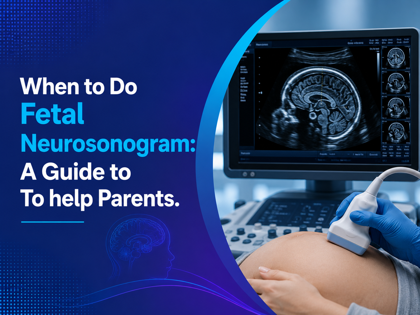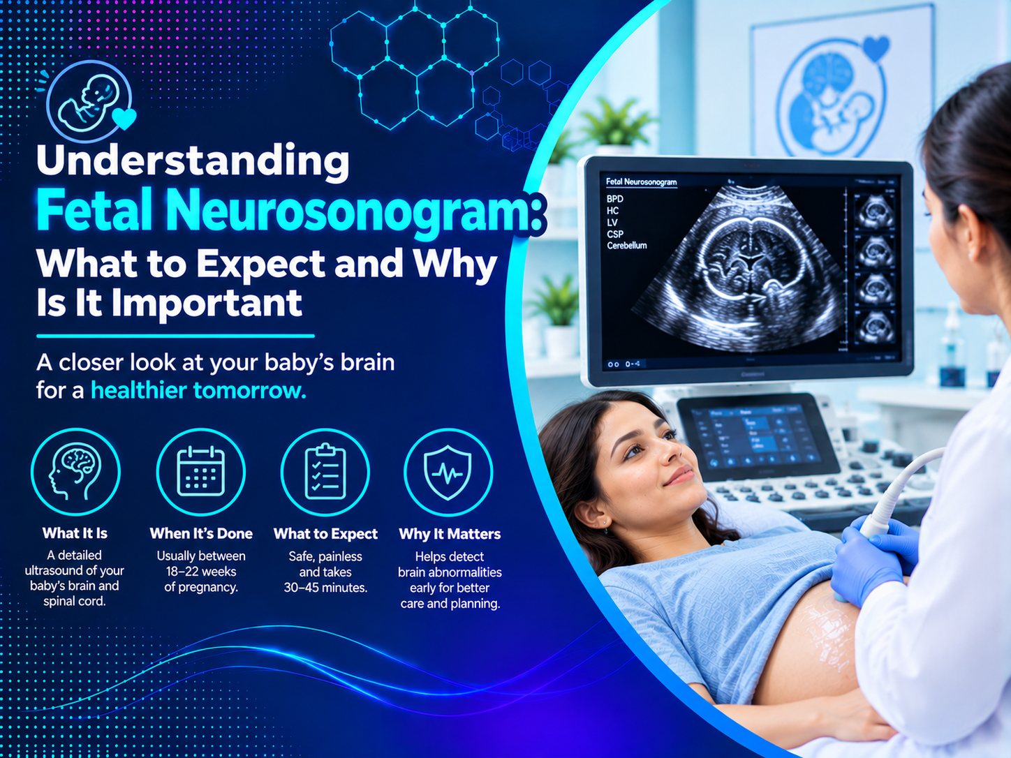
A routine pregnancy scan checks many things at once, but sometimes a doctor wants a much closer look at the baby’s brain and spine. Understanding Fetal Neurosonogram testing helps you know why this detailed scan is advised, what happens during it, and how the results guide your care. This guide explains it in simple terms so you can walk in prepared rather than worried.
What Is a Fetal Neurosonogram?
A fetal neurosonogram is a detailed ultrasound of your baby’s brain and spine. Guidelines from the International Society of Ultrasound in Obstetrics and Gynecology (ISUOG) describe it as a dedicated exam that needs specific expertise and advanced equipment. It is also called targeted neurosonography or a detailed fetal brain scan.
- Purpose: a closer, diagnostic look at the brain and spine
- Method: ultrasound with sound waves, with no radiation
- When: advised when a routine scan or a risk factor needs follow-up
How a Fetal Neurosonogram Differs From a Routine Anomaly Scan
Routine second trimester scans check the brain in three standard views, usually through the abdomen. A neurosonogram goes further, using many angles and sometimes a vaginal probe for sharper images.
| Feature | Routine Anomaly Scan | Fetal Neurosonogram |
| Purpose | Screening | Detailed diagnosis |
| Brain views | Three standard views | Multiple angles, front to back and side to side |
| Approach | Through the abdomen | Abdomen, sometimes a vaginal probe |
| Who needs it | All pregnancies | Pregnancies with a specific concern |
Why Understanding Fetal Neurosonogram Is Important
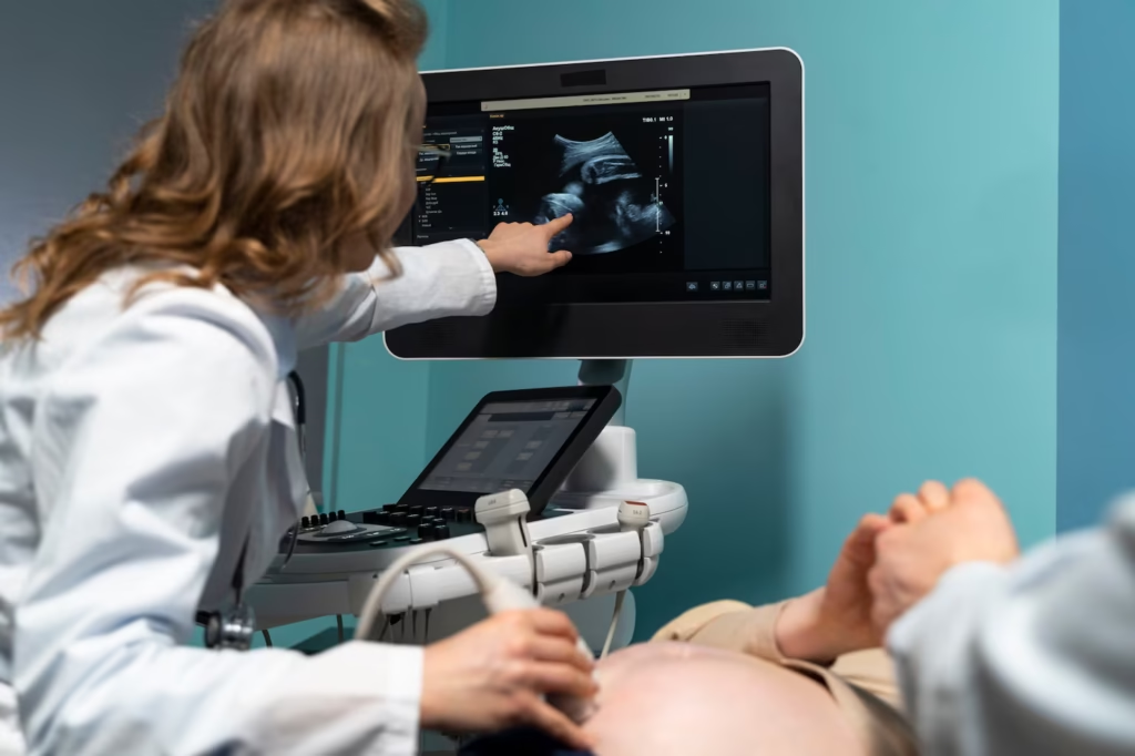
The baby’s brain keeps changing throughout pregnancy, so a closer look can reveal details that standard views may miss. ISUOG advises that fetal brain MRI should be guided by the findings of the neurosonogram, which helps avoid unnecessary extra tests.
- Confirms or rules out a concern with more certainty
- Guides decisions about further tests
- Helps doctors plan delivery and newborn care
- Gives parents clear information for counseling
- Often brings reassurance when findings are normal
What Does a Fetal Neurosonogram Examine?
The scan studies the main structures of the brain and spine, one plane at a time.
- The fluid spaces inside the brain, called ventricles
- The band of fibers connecting the two sides, called the corpus callosum
- The cerebellum, at the back of the brain
- The folds on the brain surface, which develop as pregnancy advances
- The spine, from top to bottom
When Is a Fetal Neurosonogram Recommended?
Doctors usually advise this scan for a specific reason. Common reasons include:
- A possible brain or spine finding on a routine or NT scan
- A family history, or an earlier pregnancy, with a brain or spine condition
- A heart condition found in the baby
- Monochorionic twins, who share one placenta
- A suspected infection during pregnancy
- Exposure to medicines that can affect brain development
Being referred does not mean something is wrong. Often it is a precaution.
Best Time for a Fetal Neurosonogram
Most detailed neurosonograms are done at about 20 weeks or later, after the routine anomaly scan. Because the brain changes shape week by week, timing affects what can be seen. In selected cases, specialists scan earlier, around 12 to 16 weeks, when thin skull bones allow a clear view.
If your doctor advises one, booking a Fetal Neurosonogram in Indore promptly helps you stay within the best window.
What to Expect During Your Fetal Neurosonogram
The scan is painless and similar to a routine ultrasound, though it takes longer.
- You lie down comfortably while gel is applied to your abdomen.
- The sonographer scans the baby’s head and spine from several angles.
- If the baby is head down, a vaginal probe may be used for sharper images.
- Sometimes 3D ultrasound is added to show structures more clearly.
- A specialist reviews the findings and shares a report with your doctor.
Wear comfortable clothing, bring your earlier scan reports, and allow extra time.
Tests That May Go Along With a Fetal Neurosonogram
If an infection during pregnancy is suspected, your doctor may order blood tests to check for it. A reliable Pathology Lab in Indore can process these infection and antibody panels. Some families are also offered genetic tests, while fetal MRI is considered only if the neurosonogram findings call for it.
Understanding Fetal Neurosonogram Results
Results usually fall into one of three groups.
- Normal: the brain and spine look as expected, and routine care continues.
- Minor or uncertain finding: a small variation that may need a repeat scan after a few weeks.
- Clear abnormality: your doctor refers you to a fetal medicine specialist and may add MRI, genetic tests, and counseling.
Outcomes depend on the exact finding, so ask your doctor to explain what yours means for your baby.
Fetal Neurosonogram vs Fetal MRI
Ultrasound is the main tool for checking the fetal brain, and the neurosonogram is usually the first detailed step. ISUOG notes that MRI should be advised based on the specialist’s neurosonogram findings, not on suspicion at screening alone. In practice, MRI is added only when it can give extra information about a specific finding.
- Neurosonogram: the first detailed test, showing the brain in real time
- Fetal MRI: a second-step test, used only when more detail is needed
Tips to Feel Prepared Before Your Appointment
A little preparation can ease nerves on the day.
- Write down your questions so you do not forget them
- Bring your partner or a trusted person for support
- Ask how and when you will receive your results
What Happens After the Scan
Your report goes to your obstetrician, who plans the next steps. A full-service Diagnostic Centre in Indore can often arrange repeat scans and related tests in one place, which keeps your records together.
Why Expertise and Equipment Matter
Because the exam is targeted and detailed, quality depends on the sonographer’s training and the machine’s resolution. A High-Quality Fetal Neurosonogram in Indore relies on three things: an experienced fetal imaging specialist, high-resolution equipment with vaginal and 3D options, and a clear written report.
Choosing Where to Get Scanned
Look for a centre that pairs fetal imaging experience with clear, supportive communication. Ultrascan Diagnostic Centre offers pregnancy imaging including early anomaly scans, growth scans, fetal echocardiography, and 3D/4D targeted imaging, so related scans can be done in one place. Parents searching for the best diagnostic centre in Indore can ask the team directly about their neurosonogram process.
Frequently Asked Questions
1. Is a fetal neurosonogram the same as an anomaly scan?
No. The anomaly scan is routine screening for all pregnancies, while a neurosonogram is a detailed scan of the brain and spine done for a specific reason.
2. Is a fetal neurosonogram safe?
Yes. It uses sound waves, not radiation, and a vaginal probe can be used safely when sharper images are needed.
3. Does a referral mean my baby has a problem?
No. Referrals often happen because of a family history, a twin pregnancy, or a small finding that needs a closer look.
4. When is a fetal neurosonogram done?
Usually at about 20 weeks or later, though specialists may scan earlier in selected cases.
5. Will I need an MRI afterward?
Only if the neurosonogram findings suggest it. Your doctor will decide based on what the specialist sees.
Conclusion
Understanding Fetal Neurosonogram testing takes much of the fear out of a scan that can sound intimidating. It is a careful, detailed look at your baby’s brain and spine, and it gives your doctor the information needed to plan the right care.
Book your detailed fetal scan consultation at Ultrascan Diagnostic Centre today. Call ahead or walk in to plan your appointment. This article is for general information and does not replace advice from your gynecologist.