Thyroid problems are among the most common and most commonly missed health conditions in the world. If you have been experiencing symptoms such as persistent fatigue, unexplained weight changes, a lump in the neck, hair thinning, or mood disturbances, your doctor may have suggested investigating your thyroid. And that immediately raises the question many people ask: Should I get a thyroid ultrasound or just a blood test for thyroid problems?
The answer, in short, is that both serve different and equally important purposes, and in many cases, you will need both. A thyroid blood test tells you how the thyroid is functioning. A thyroid ultrasound tells you what the thyroid looks like. These are two distinct pieces of information, and neither one alone gives the complete picture.
At Ultrascan Diagnostics, your trusted ultrasound diagnostic centre in Indore, we perform high-resolution thyroid ultrasound scans that complement blood test findings and give your doctor the structural detail needed for accurate diagnosis. This guide explains both tests clearly, what they show, when each is needed, and why combining them is often the smartest approach.
What Does a Thyroid Blood Test Show – And What Are Its Limits?
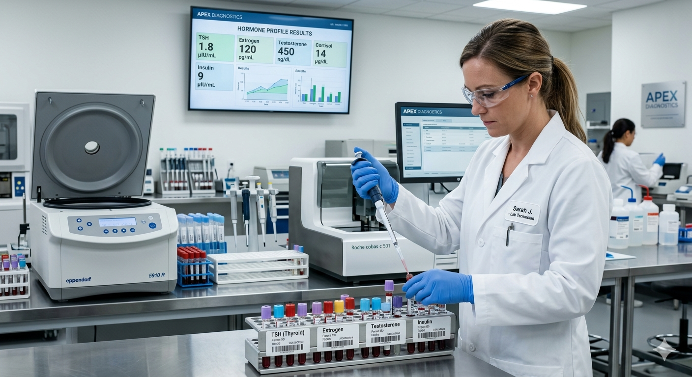
A thyroid blood test measures the levels of hormones and antibodies in your blood that reflect how well your thyroid gland is working. The most commonly ordered tests include:
• TSH (Thyroid-Stimulating Hormone): The primary screening test. TSH is produced by the pituitary gland and tells the thyroid how much hormone to make. A high TSH suggests the thyroid is underactive (hypothyroidism); a low TSH suggests it is overactive (hyperthyroidism).
• Free T3 and Free T4: The actual hormones produced by the thyroid itself. Measuring these alongside TSH provides a fuller picture of thyroid function.
• Anti-TPO and Anti-Thyroglobulin Antibodies: These detect autoimmune thyroid disease – such as Hashimoto’s thyroiditis (the most common cause of hypothyroidism) or Graves’ disease (the most common cause of hyperthyroidism).
• Thyroglobulin: Used as a marker after thyroid cancer treatment to check for recurrence.
• Calcitonin: Sometimes tested when medullary thyroid cancer is suspected.
Thyroid blood tests are excellent at answering one question: is the thyroid producing the right amount of hormone? They are the foundation of thyroid diagnosis and the primary tool for monitoring patients on thyroid medication.
However, thyroid blood tests have important limitations:
• They cannot detect structural abnormalities within the thyroid gland, such as nodules, cysts, or goitre.
• They cannot distinguish between a simple benign nodule and a potentially malignant one.
• A completely normal TSH result does not rule out the presence of thyroid nodules or early thyroid cancer – because many thyroid cancers do not affect hormone production at all.
• They cannot assess the size, vascularity, or texture of the thyroid gland.
• They give no information about whether a nodule has calcifications, irregular borders, or other features associated with malignancy.
What Does a Thyroid Ultrasound Scan Show That a Blood Test Cannot?
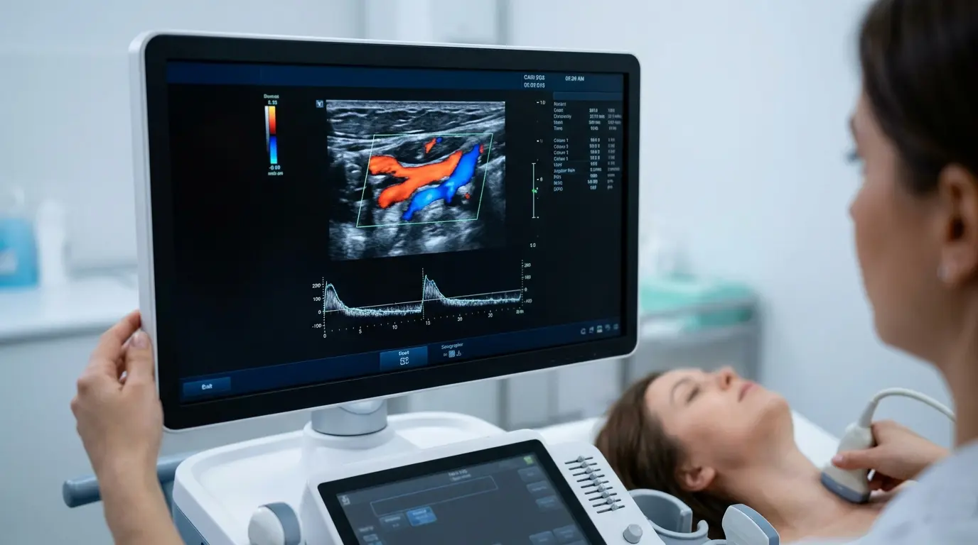
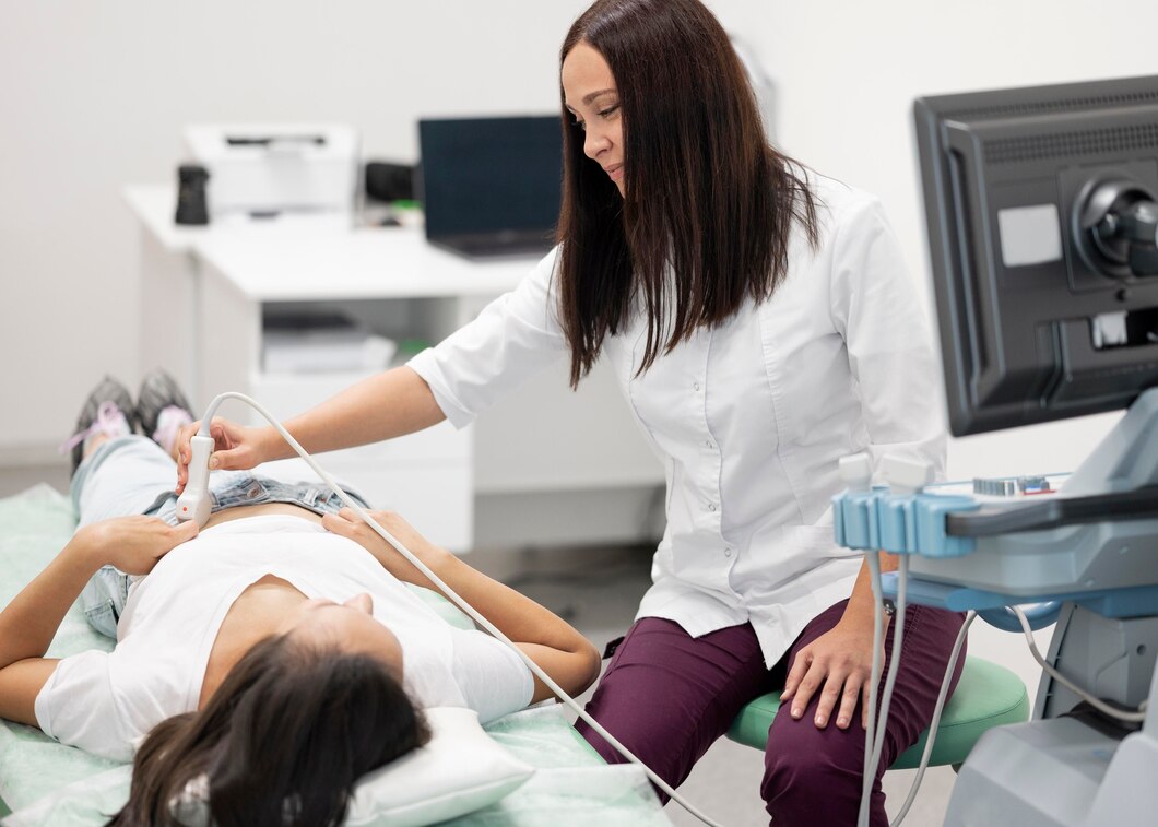
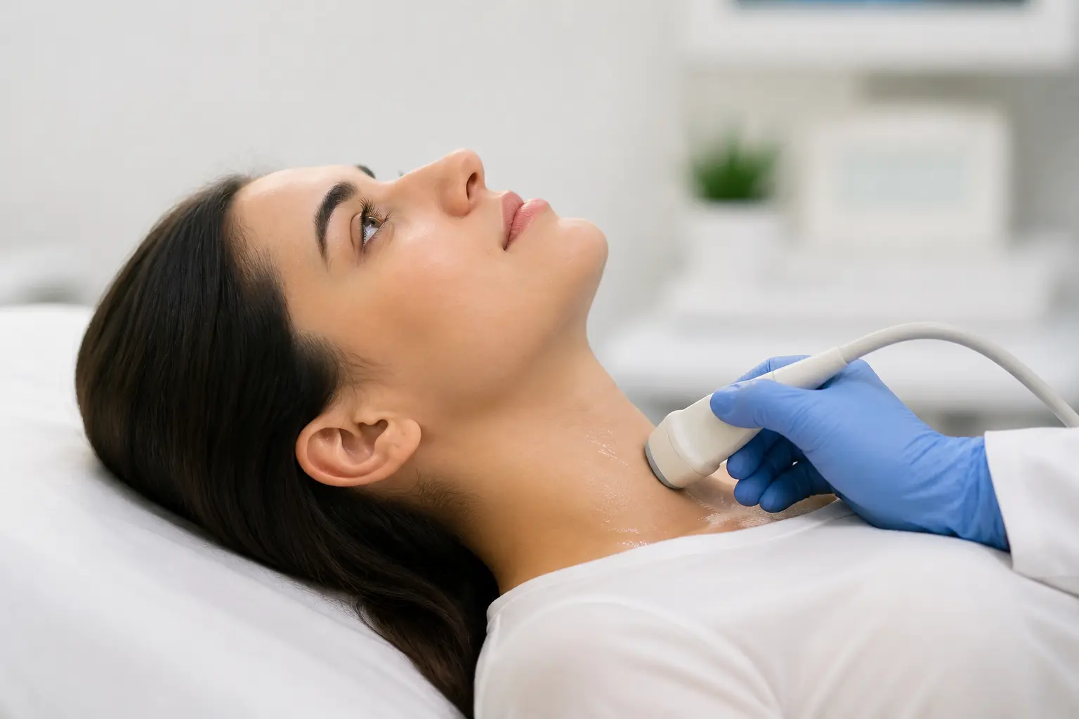
A thyroid ultrasound scan uses high-frequency sound waves to create detailed, real-time images of the thyroid gland. It is the gold-standard imaging tool for thyroid structural assessment – completely safe, radiation-free, and capable of detecting abnormalities as small as 2 to 3 millimetres.
Here is what a thyroid ultrasound can reveal:
1. Thyroid Nodules – Their Number, Size, and Characteristics
Thyroid nodules are extremely common, affecting up to 50% of the population when screened by ultrasound, though most are benign. An ultrasound precisely measures each nodule, records its location, and assesses features that indicate its risk level:
• Solid vs fluid-filled (cystic) – solid nodules carry a higher risk of malignancy
• Irregular or ill-defined margins – associated with higher risk
• Micro-calcifications – tiny calcium deposits linked to papillary thyroid cancer
• Taller-than-wide shape – a suspicious feature on ultrasound
• Increased or chaotic internal blood flow on Colour Doppler – another risk indicator
• Hypoechoic texture (darker than surrounding tissue) raises concern for malignancy
Based on these features, radiologists assign nodules a risk category using standardised systems – helping doctors decide whether a nodule needs a biopsy (FNAC – Fine Needle Aspiration Cytology), repeat monitoring, or simple reassurance.
2. Goitre – Thyroid Gland Enlargement
A goitre is an enlargement of the thyroid gland. It can be diffuse (the whole gland is enlarged) or nodular (enlarged due to one or more nodules). A thyroid ultrasound precisely measures the gland’s dimensions and estimates its total volume – information that is essential for monitoring growth over time and for planning treatment such as medication, radioiodine therapy, or surgery.
3. Thyroiditis – Inflammation of the Thyroid
Both Hashimoto’s thyroiditis and De Quervain’s thyroiditis (a painful, usually temporary thyroid inflammation) produce characteristic changes in the appearance of the thyroid gland on ultrasound. In Hashimoto’s thyroiditis, the gland typically appears heterogeneous (uneven texture), smaller than normal over time, and may show a characteristic coarsened echotexture. These features, combined with positive antibody blood tests, confirm the diagnosis.
4. Thyroid Cysts
Pure thyroid cysts – smooth, fluid-filled sacs – are almost always benign. An ultrasound can immediately distinguish a simple cyst from a complex cystic nodule (which has solid components and requires closer evaluation). This distinction saves patients from unnecessary anxiety and invasive procedures.
5. Lymph Node Assessment
During a thyroid ultrasound, the radiologist also assesses nearby lymph nodes in the neck. Suspicious lymph nodes – those that are enlarged, rounded, or show internal calcifications – may indicate spread of thyroid cancer and will be flagged for further investigation. This contextual assessment is simply not possible with a blood test.
Should I Get a Thyroid Ultrasound or Just a Blood Test? A Practical Guide
The clearest way to answer should I get a thyroid ultrasound or just a blood test for thyroid problems is to match the right test to the right clinical question.
Get a Thyroid Blood Test First If:
• You are experiencing symptoms of hypothyroidism – fatigue, weight gain, cold intolerance, dry skin, constipation, hair loss, depression, or slow heart rate
• You are experiencing symptoms of hyperthyroidism – weight loss, rapid heartbeat, anxiety, tremor, heat intolerance, or diarrhoea
• You are monitoring a known thyroid condition and are already on medication
• Your doctor is screening for autoimmune thyroid disease due to family history or other autoimmune conditions
• You are pregnant or planning to become pregnant, as thyroid function is critical in early pregnancy
Get a Thyroid Ultrasound Scan If:
• Your doctor can feel a lump or nodule in your neck during examination
• You can feel or see a swelling at the base of your throat
• A nodule or abnormality has been noticed incidentally on another scan (such as a neck CT or carotid Doppler)
• You have a family history of thyroid cancer – especially medullary thyroid cancer or MEN syndrome
• You have had previous radiation to the head, neck, or chest
• Your blood tests are abnormal and the doctor wants to understand the structural cause
• You have a known thyroid condition and your doctor wants to monitor the gland’s size or any nodules over time
• You have hoarseness, difficulty swallowing, or a sensation of pressure in the throat without a clear cause
Get Both Together If:
• You have both functional symptoms (fatigue, weight changes) AND a palpable neck lump
• You have a newly discovered thyroid nodule that needs both structural characterisation and functional context
• Your doctor is planning a biopsy and needs both sets of information before proceeding
• You are having a comprehensive thyroid health review after diagnosis of thyroid disease
When Is a Thyroid Ultrasound Used to Guide a Biopsy (FNAC)?
If a thyroid nodule has features on ultrasound that suggest it may be malignant, the next step is often a Fine Needle Aspiration Cytology (FNAC) – a minimally invasive biopsy where a thin needle is used to extract a small sample of cells from the nodule for laboratory analysis.
At Ultrascan Diagnostics, we perform ultrasound-guided FNAC – where the ultrasound probe is used in real time to guide the needle precisely into the nodule being sampled. This significantly improves accuracy, reduces the risk of sampling the wrong area, and increases the chance of obtaining a diagnostic result on the first attempt.
Ultrasound-guided FNAC is recommended when a nodule is:
• Solid and hypoechoic with any suspicious features
• Larger than 1 centimetre with intermediate-risk features
• Growing on serial ultrasound monitoring
• In a patient with a personal or family history of thyroid cancer
• Associated with suspicious lymph nodes on the same scan
The procedure takes only a few minutes, requires no anaesthesia, and the patient can return to normal activities the same day. Cytology results are typically available within a few days and allow your doctor to plan the most appropriate treatment.
What to Expect During a Thyroid Ultrasound Scan at Ultrascan Diagnostics
A thyroid ultrasound at our ultrasound scan in Indore centre is quick, comfortable, and completely painless. Here is what happens:
• No preparation needed: You do not need to fast, and no special preparation is required before a thyroid ultrasound.
• Positioning: You will lie on your back with a small pillow or rolled towel placed under your neck to gently extend it, giving the radiologist a clear view of the thyroid.
• The scan: A water-based gel is applied to the front of the neck and a small probe is moved gently across the skin. The radiologist examines the entire thyroid gland, both lobes, the isthmus, and the surrounding lymph nodes.
• Duration: A standard thyroid ultrasound takes approximately 15 to 20 minutes. If a Colour Doppler assessment or FNAC is being performed, allow an additional 10 to 20 minutes.
• Results: A detailed written report with ultrasound images is generated and provided to your referring doctor, usually on the same day or within 24 hours.
Why Patients Across Indore Choose Ultrascan Diagnostics for Thyroid Ultrasound
When it comes to thyroid health, the quality of your ultrasound diagnostic imaging directly affects the quality of your diagnosis. Here is what sets Ultrascan Diagnostics apart:
• High-resolution ultrasound machines: Our equipment detects nodules as small as 2mm – critical for early detection of thyroid abnormalities.
• Colour Doppler capability: We assess the blood flow characteristics of thyroid nodules and the gland as a whole – an additional layer of diagnostic information.
• Ultrasound-guided FNAC: Precise, minimally invasive biopsy of thyroid nodules performed by experienced hands – on-site, without the need for a separate referral.
• Fast, same-day reports: Your endocrinologist or physician receives the information they need quickly – reducing the time between scan and next clinical decision.
• Affordable, transparent pricing: No hidden fees. We offer competitive packages for thyroid ultrasound, with or without Colour Doppler and FNAC.
• Easy to reach: Located on Pipliyahana Road, Indore – easily accessible for patients from Vijay Nagar, Scheme 54, Bicholi Mardana, and surrounding areas.
Frequently Asked Questions: Thyroid Ultrasound vs Blood Test
Q1. Can a thyroid ultrasound detect cancer that a blood test misses?
Yes – and this is one of the most important reasons to understand why you should not rely on blood tests alone. The majority of thyroid cancers – particularly papillary thyroid cancer, which is the most common type – do not significantly affect TSH, T3, or T4 levels. A person with early thyroid cancer may have completely normal thyroid function tests while harbouring a malignant nodule in the gland. A thyroid ultrasound, on the other hand, can detect suspicious nodule features – such as micro-calcifications, irregular margins, increased vascularity, and a taller-than-wide orientation – that prompt further investigation through FNAC. This is why ultrasound is recommended for all palpable thyroid abnormalities, even when blood tests are normal.
Q2. How often should I have a thyroid ultrasound if I have nodules?
The follow-up frequency for thyroid nodules depends on their size and ultrasound risk features, as well as the results of any biopsy performed. For low-risk or very low-risk nodules (those with reassuring features on ultrasound and benign FNAC results), a repeat scan at 12 to 24 months is typically recommended to confirm stability. If a nodule grows by more than 20% in volume or develops new suspicious features on follow-up, re-evaluation with FNAC may be necessary. For intermediate- or high-risk nodules, closer monitoring – every 6 to 12 months – may be advised. Your endocrinologist or radiologist will provide a personalised monitoring plan based on your specific ultrasound findings. Ultrasound indore services at Ultrascan Diagnostics include structured follow-up scheduling to ensure your nodules are tracked consistently over time.
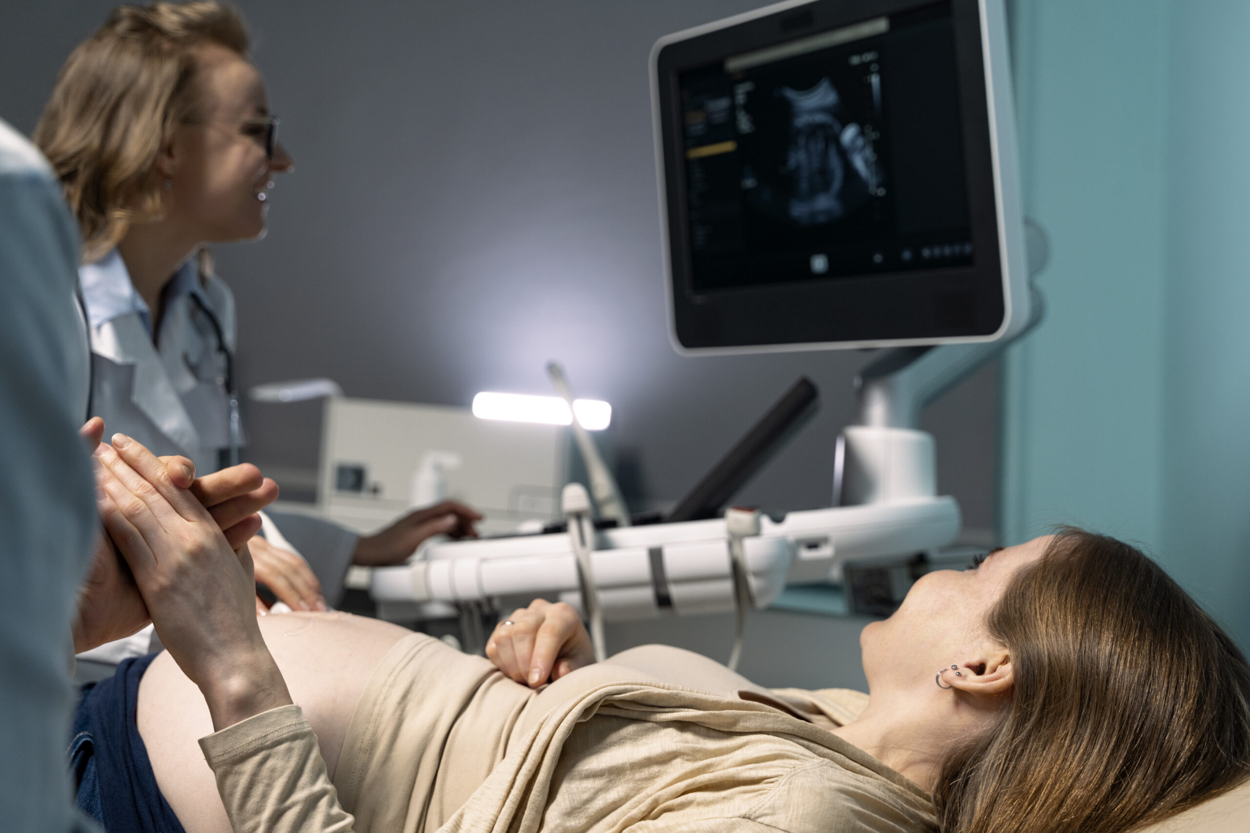
Q3. Is a thyroid ultrasound safe during pregnancy?
Yes – a thyroid ultrasound is completely safe during pregnancy and can be performed at any stage. It uses sound waves, not radiation, making it entirely risk-free for both the mother and the developing baby. Thyroid health is particularly important during pregnancy because thyroid hormones are essential for the baby’s neurological development, especially in the first trimester before the baby’s own thyroid becomes functional. If a pregnant woman is found to have a thyroid nodule – whether through routine examination or incidentally on an obstetric scan – a thyroid ultrasound is the recommended and safest way to evaluate it. Many endocrinologists recommend that pregnant women with known thyroid nodules have at least one thyroid ultrasound during pregnancy to monitor for significant changes.