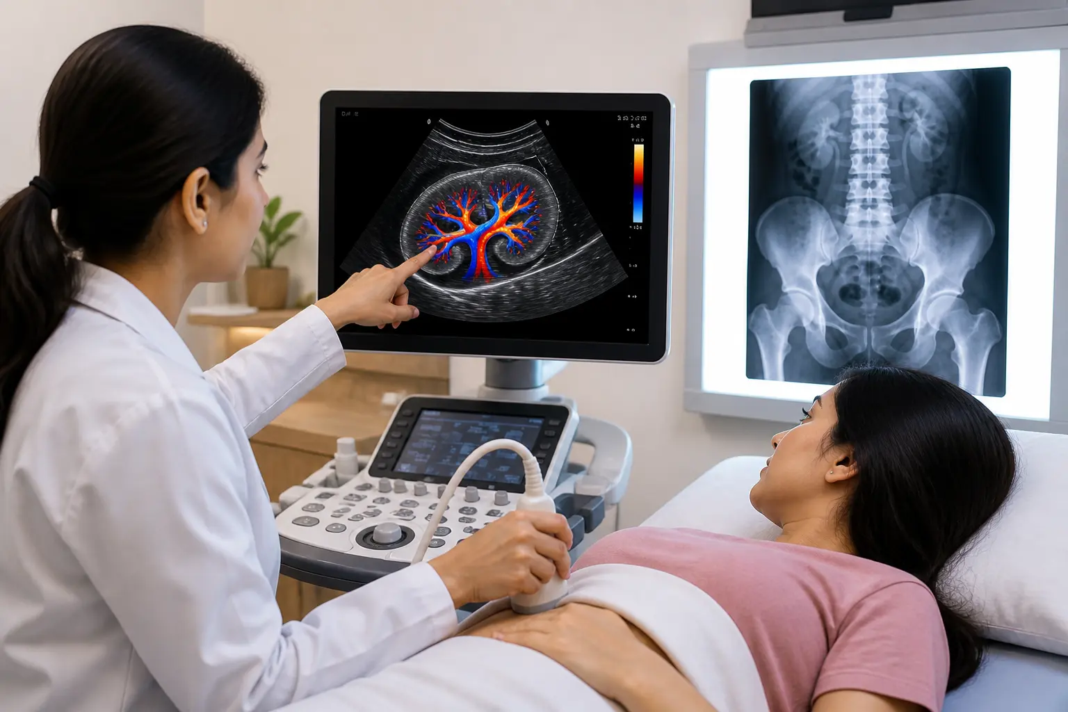
Arthritis often develops gradually, with joint inflammation sometimes present well before pain or swelling becomes obvious. MSK Ultrasound Enhances Arthritis Diagnosis by giving doctors a real-time, detailed view of what is actually happening inside a joint, revealing changes that a physical exam alone might miss. This guide explains how this imaging technique works, what it can detect, and why it has become such a valuable tool for both diagnosing and tracking arthritis over time.
What Is Musculoskeletal Ultrasound?
Musculoskeletal ultrasound, often shortened to MSK ultrasound, uses sound waves to create detailed, real-time images of joints, tendons, muscles, and surrounding soft tissue. Unlike X-rays, which primarily show bone, this technique captures the soft tissue changes that are often the earliest signs of inflammatory joint disease.
Because the images update live as the joint moves, doctors can observe structures dynamically rather than relying on a single static picture. This real-time capability is a major reason MSK Ultrasound Enhances Arthritis Diagnosis so effectively compared to imaging methods that only capture a fixed moment in time.
Why Early Arthritis Detection Is So Challenging
Arthritis, particularly rheumatoid arthritis, can be difficult to diagnose in its earliest stages because symptoms often overlap with other conditions and inflammation may not yet be severe enough to detect through a physical exam alone. Blood tests for markers like rheumatoid factor can also come back normal in some patients who genuinely have the disease.
This diagnostic gap is precisely where musculoskeletal ultrasound has proven especially valuable, since research shows it can detect synovitis, the joint inflammation at the heart of rheumatoid arthritis, with notably higher accuracy than clinical examination alone. Catching this inflammation early allows doctors to begin treatment before significant joint damage has a chance to develop.
What MSK Ultrasound Can Detect in Arthritis
This imaging technique reveals several specific changes associated with inflammatory joint disease, each offering valuable diagnostic information.
- Synovitis, thickening of the joint lining that signals active inflammation
- Joint effusion, excess fluid buildup within the joint space
- Bone erosions, early structural damage that may not yet be visible on standard X-rays
- Increased blood flow, detected using a specialized Doppler setting, indicating active inflammatory activity
- Tenosynovitis, inflammation of the tendon sheath, common in several forms of inflammatory arthritis
Detecting these changes early, sometimes even before a joint appears visibly swollen, gives doctors a meaningful head start in managing the condition before it progresses.
How MSK Ultrasound Compares to Other Imaging Methods
Different imaging techniques each offer distinct advantages when evaluating arthritis, and doctors often choose based on what specific information is needed.
| Imaging Method | Strengths | Limitations |
| X-ray | Shows established bone damage, widely available | Cannot detect early soft tissue inflammation |
| MSK Ultrasound | Real-time, detects early synovitis and effusion, no radiation | Operator-dependent, limited depth penetration |
| MRI | Highly detailed, excellent soft tissue contrast | Expensive, time-consuming, less accessible |
Ultrasound’s combination of accessibility, affordability, and strong sensitivity for early inflammation makes it a particularly practical choice for both initial diagnosis and ongoing monitoring.
Using MSK Ultrasound to Monitor Arthritis Treatment Response
Beyond initial diagnosis, this imaging technique plays an increasingly important role in tracking how well arthritis treatment is working over time. Doctors can repeat scans at intervals to measure whether synovial thickening, fluid buildup, or abnormal blood flow is improving, stable, or worsening under a given treatment plan.
This ongoing monitoring is particularly valuable because some patients can appear clinically well, with no visible swelling, while scans still show subclinical inflammation that carries a real risk of future joint damage. Identifying this hidden activity allows doctors to adjust treatment proactively rather than waiting for a visible flare to occur.
Which Joints Are Commonly Examined?
MSK ultrasound can be used to evaluate nearly any joint, though certain areas are examined more frequently due to how commonly they are affected by arthritis.
- Wrists and hands, frequently affected in rheumatoid arthritis, particularly the small finger joints
- Knees, commonly evaluated for effusion and synovitis across multiple arthritis types
- Shoulders, assessed for both joint inflammation and associated tendon involvement
- Ankles and feet, particularly relevant in conditions like gout and psoriatic arthritis
- Elbows, evaluated when symptoms suggest inflammatory involvement in this joint
Differentiating Between Types of Arthritis
One particularly useful application of this imaging technique is helping distinguish between different forms of inflammatory arthritis, which can sometimes present with overlapping symptoms. Gout, for example, often shows a distinctive pattern on ultrasound involving crystal deposits, while rheumatoid arthritis typically presents with a more symmetric pattern of synovial thickening across multiple joints.
This diagnostic clarity matters significantly, since treatment approaches for gout, rheumatoid arthritis, and psoriatic arthritis differ meaningfully from one another. Identifying the correct underlying cause earlier helps doctors choose the right treatment path from the start rather than relying on trial and error.
What to Expect During an MSK Ultrasound Examination

Understanding the process can help ease any uncertainty before your appointment.
- No special preparation, such as fasting, is required before this type of scan
- A gel is applied to the skin over the joint being examined
- The sonographer moves the transducer over the area, sometimes asking you to move the joint during the scan
- A Doppler setting may be used briefly to assess blood flow within the joint
- The scan is painless and typically takes 15 to 30 minutes, depending on how many joints are being examined
Finding the Right Centre for Joint Imaging
Since this type of imaging requires specific technical skill, choosing an experienced facility genuinely affects the accuracy of your results. Patients experiencing unexplained joint pain often search for Ultrasound for muscle pain and joint symptoms as a starting point before learning that a more specialized musculoskeletal scan may better address their specific concern.
Specialized Imaging for Joint and Soft Tissue Concerns
For patients specifically seeking Musculoskeletal ultrasound in Indore, it is worth confirming that the facility has sonographers trained specifically in joint and soft tissue imaging, since this differs meaningfully from general abdominal or obstetric scanning experience.
Comprehensive MSK Imaging Services
Patients researching MSK ultrasound in Indore for suspected arthritis or persistent joint symptoms should look for a centre offering both grayscale and Doppler imaging, since combining these views provides the most complete diagnostic picture.
Why Choose Ultrascan Diagnostic Centre
Ultrascan Diagnostic Centre is recognized as one of the best diagnostic centre in Indore for musculoskeletal imaging, combining modern ultrasound technology with sonographers experienced in joint-specific scanning protocols.
A Trusted Partner for Ongoing Joint Monitoring
As an established Diagnostic Centre in Indore, Ultrascan supports patients through both initial arthritis diagnosis and the ongoing monitoring needed to track treatment response over time.
Frequently Asked Questions
1. Can MSK ultrasound diagnose rheumatoid arthritis on its own?
MSK ultrasound is a powerful supporting tool, but diagnosis typically combines imaging findings with blood tests and a clinical examination. It is especially useful for detecting synovitis, even in cases where blood markers appear normal.
2. Is MSK ultrasound better than an X-ray for arthritis?
Each has its own strengths, since X-rays are better at showing established bone damage, while MSK ultrasound excels at detecting early soft tissue inflammation that X-rays cannot capture. Many doctors use both together for a more complete evaluation.
3. How often should arthritis patients get an MSK ultrasound for monitoring?
This depends on disease activity and treatment response, but many doctors recommend periodic scans every few months during active treatment adjustments. Your rheumatologist will determine the right monitoring schedule based on your specific condition.
4. Does MSK ultrasound involve any radiation?
No, this technique uses sound waves rather than radiation, making it safe for repeated use during ongoing arthritis monitoring. This safety profile is one of the key reasons it has become so widely used for tracking treatment response over time.
5. Can MSK ultrasound tell the difference between gout and rheumatoid arthritis?
Yes, these conditions often show distinct patterns on ultrasound, such as crystal deposits in gout versus symmetric synovial thickening in rheumatoid arthritis. This distinction helps doctors choose the right treatment approach more confidently and quickly.
Conclusion
MSK Ultrasound Enhances Arthritis Diagnosis by revealing joint inflammation long before it becomes visibly obvious, giving doctors the information needed to intervene earlier and more effectively. Whether used for initial diagnosis or tracking how well a treatment plan is working, this imaging technique has become an indispensable tool in modern rheumatology care.
Book your musculoskeletal ultrasound today at Ultrascan Diagnostic Centre and get clear, detailed answers about your joint symptoms. Walk in or call ahead to schedule your appointment and take a confident step toward better joint health.