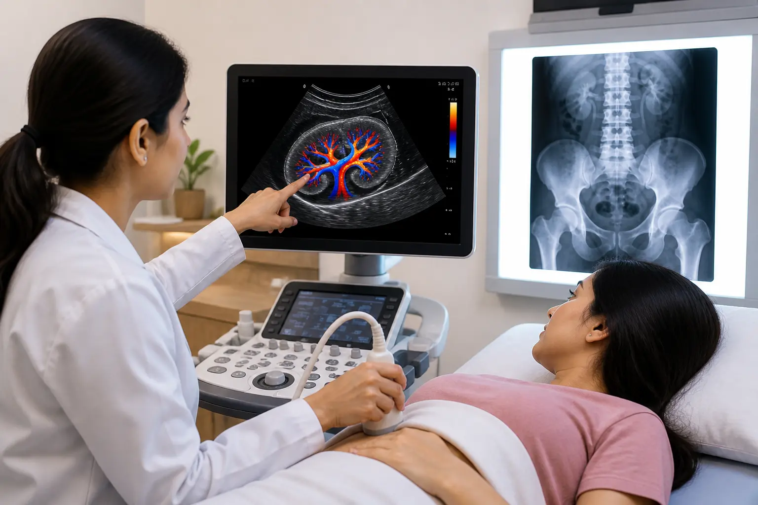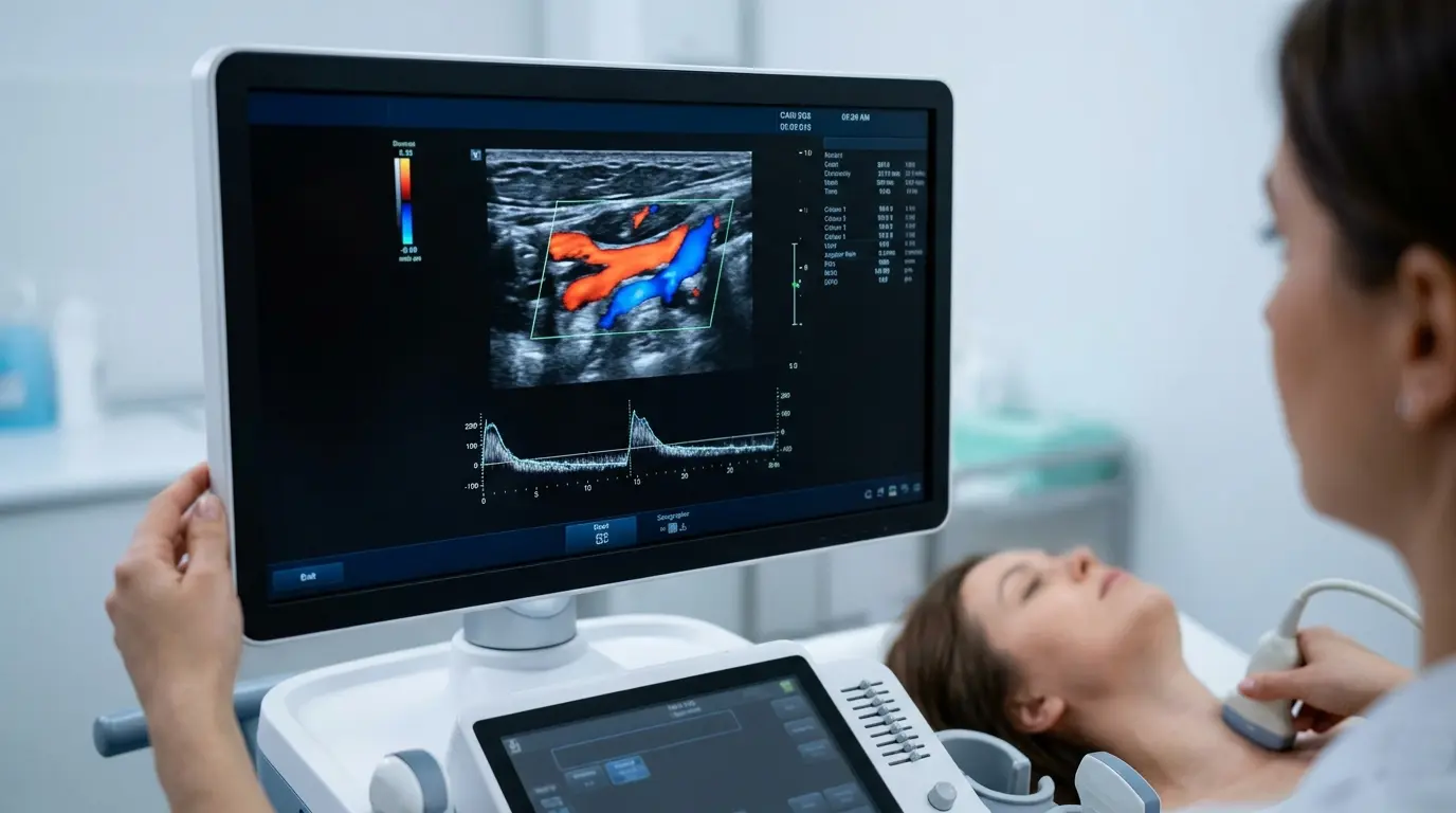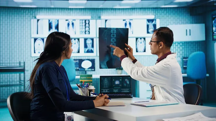
X-rays are often the first imaging test a doctor orders, and for good reason. The Diagnostic Power of X-Rays lies in how quickly and clearly they reveal what is happening inside the body, from a simple fracture to signs of serious lung disease. This guide explores exactly what X-rays can detect, how the technology works, and why this decades-old tool remains one of the most valuable resources in modern diagnostics.
How X-Rays Work to Reveal What’s Inside the Body
X-rays are a form of electromagnetic radiation that passes through the body and is absorbed differently by different tissues. Dense structures like bone absorb more radiation and appear white on the image, while softer tissues absorb less and appear in shades of gray, creating a clear contrast that doctors use to spot abnormalities.
This simple principle is what gives X-rays their diagnostic strength, since the resulting image reveals structural details that would otherwise be invisible without surgery. Understanding this basic science helps explain why the Diagnostic Power of X-Rays remains so valuable despite the availability of newer, more advanced imaging technologies.
Bone and Joint Conditions X-Rays Can Detect
Bone imaging remains one of the most well-known uses of X-ray technology, and for good reason, since bones show up with exceptional clarity on these scans.
- Fractures and breaks, including hairline fractures that are not visible externally
- Dislocations, where a joint has shifted out of its normal position
- Osteoarthritis, identified through narrowed joint space and bone spurs
- Osteoporosis, which shows as reduced bone density on imaging
- Bone tumors, both benign growths and more concerning malignant lesions
- Bone infections, such as osteomyelitis, which can alter bone structure over time
Because bones absorb X-ray radiation so effectively, these conditions are often visible with a high degree of clarity, making X-rays the natural first choice for most bone-related concerns. Getting these scans read correctly matters just as much as the scan itself, which is why patients often seek out the best diagnostic centre in Indore when a fracture or joint issue needs a fast, accurate answer.
Chest and Lung Conditions Detected Through X-Ray Imaging
A chest X-ray is one of the most frequently ordered scans in medicine, largely because it can reveal so much about the heart and lungs in a single image.
Respiratory Conditions
Chest X-rays are commonly used to detect pneumonia, tuberculosis, and chronic obstructive pulmonary disease, all of which produce visible changes in lung tissue. Doctors also rely on chest imaging to identify lung cancer, particularly when a suspicious mass or nodule needs further evaluation.
Heart-Related Findings
While X-rays are not the primary tool for detailed heart imaging, they can reveal an enlarged heart, a condition called cardiomegaly, which often points toward underlying heart failure. This makes chest X-rays a useful early indicator even before more specialized cardiac testing is ordered.
Fluid and Air Abnormalities
Conditions such as pulmonary edema, where fluid builds up in the lungs, and pneumothorax, where air collects abnormally around the lung, both show up clearly on a standard chest X-ray. These findings often guide urgent treatment decisions in emergency settings.
Abdominal Conditions X-Rays Can Help Identify
Beyond bones and lungs, X-ray imaging also plays a role in evaluating certain abdominal conditions, though it is often paired with other tests for a complete picture.
- Intestinal obstructions, which show as abnormal gas patterns in the bowel
- Kidney stones, particularly larger stones that are visible on standard imaging
- Gallstones, in cases where the stones contain enough calcium to appear on X-ray
- Perforated organs, identified through free air visible inside the abdominal cavity
- Swallowed foreign objects, especially in children, where location needs to be confirmed quickly
Abdominal symptoms often need more than imaging alone to reach a clear diagnosis, so doctors frequently pair an X-ray with blood work from a trusted Pathology Lab in Indore to rule out infection or organ dysfunction alongside the imaging findings.
X-Ray Detection Summary by Body Area
| Body Area | Common Conditions Detected |
| Bones and joints | Fractures, osteoporosis, arthritis, bone tumors |
| Chest and lungs | Pneumonia, tuberculosis, lung cancer, COPD |
| Heart | Cardiomegaly and related structural changes |
| Abdomen | Kidney stones, intestinal obstruction, perforation |
| Dental | Tooth decay, impacted teeth, jaw abnormalities |
Why X-Rays Remain a First-Line Diagnostic Tool
Despite the growing availability of advanced imaging like MRI and CT scans, X-rays continue to hold a critical place in diagnostic medicine. Their speed, affordability, and wide availability make them the practical first step for identifying a broad range of conditions before deciding whether further imaging is needed.
A modern Digital X-Ray in Indore takes this advantage even further, using digital sensors instead of traditional film to produce sharper images with lower radiation exposure. This technology also allows results to be reviewed almost instantly, which speeds up diagnosis considerably compared to older film-based systems.
What Happens During an X-Ray Scan
Understanding the process can ease any first-time nerves about getting an X-ray done.
- You will be asked to remove any metal objects or jewelry near the area being scanned.
- A technician positions your body for the correct angle, which may involve standing, sitting, or lying down.
- You will need to hold very still for a few seconds while the image is captured.
- Multiple images from different angles may be taken for a complete view.
- A radiologist reviews the images and prepares a detailed report for your doctor.
Most X-rays involve very low radiation exposure and are considered safe for occasional diagnostic use as recommended by your doctor. Some routine Health Packages in Indore even include a basic chest X-ray as part of an annual wellness screening, particularly for smokers or older adults.
Are X-Rays Always the Right Choice?
While X-rays are powerful for many conditions, they are not always the most appropriate imaging test. Soft tissue structures like ligaments, tendons, and internal organs are often better evaluated using MRI or ultrasound, since X-rays are primarily suited to dense structures like bone.
Your doctor will decide whether an X-ray, MRI, CT scan, or ultrasound is the right starting point based on your specific symptoms and the body part being examined. In many cases, an X-ray remains the fastest and most cost-effective way to rule out or confirm the most likely explanation before considering further tests, and a well-equipped Diagnostic Centre in Indore can usually offer several of these imaging options under one roof.
Getting an Accurate X-Ray Scan in Indore
Choosing the right facility significantly affects how clearly a condition can be identified, since image quality depends heavily on both equipment and technique. Anyone searching for a reliable x ray scan in Indore should look for a centre that combines modern digital imaging with experienced radiologists who can interpret results accurately.
Frequently Asked Questions
1. Can an X-ray detect cancer?
X-rays can help identify suspicious masses or nodules, particularly in the lungs or bones, which may prompt further testing. However, a definitive cancer diagnosis usually requires additional imaging or a biopsy to confirm the finding.
2. How much radiation exposure is involved in a standard X-ray?
A single X-ray involves a very low dose of radiation, generally considered safe for occasional diagnostic use. Modern digital equipment has further reduced radiation exposure compared to older film-based X-ray machines.
3. Can X-rays detect soft tissue injuries like ligament tears?
No, X-rays are best suited to dense structures like bone and are not effective at showing soft tissue detail. If a ligament or tendon injury is suspected, your doctor will likely recommend an MRI in addition to or instead of an X-ray.
4. How quickly are X-ray results available?
With digital imaging, results are often available almost immediately for the radiologist to review, and most reports are shared with your doctor the same day. This quick turnaround makes X-rays especially useful in urgent or emergency situations.
5. Is a chest X-ray enough to rule out lung disease completely?
A chest X-ray is a strong first step, but it cannot rule out every possible lung condition on its own. If symptoms persist despite a normal X-ray, your doctor may recommend a CT scan for a more detailed evaluation.
Conclusion
The Diagnostic Power of X-Rays lies in their ability to reveal a remarkably wide range of conditions, from broken bones to lung disease, using a test that is fast, affordable, and widely available. While newer imaging technologies continue to advance, X-rays remain an essential first step in identifying what is really happening inside the body.
Book your X-ray scan today at Ultrascan Diagnostic Centre, a trusted name for accurate imaging in Indore. Walk in or call ahead to schedule your appointment and get clear, reliable answers about your health.