A baby’s heart begins forming and beating within the first few weeks of pregnancy, making it one of the earliest and most complex organs to develop. Colour Doppler Sonography has become one of the most valuable tools available for examining this development in detail, allowing doctors to see not just the structure of the heart but the actual movement of blood flowing through it. This guide explains how this technology works, why it plays such an important role in detecting congenital heart defects, and what expecting parents should know about this specialized form of screening.
What Is Colour Doppler Sonography?
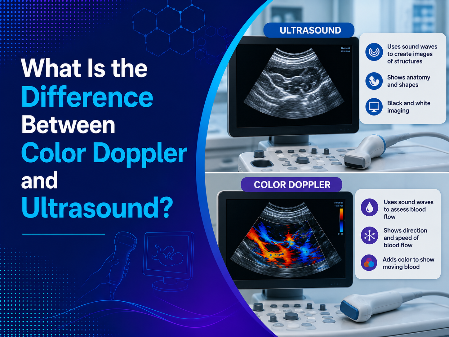
Standard ultrasound imaging shows the structure of an organ, but it cannot show how blood is actually moving through it. Colour Doppler Sonography solves this limitation by measuring the speed and direction of blood flow, then displaying this information as color overlaid on the standard black-and-white image.
Blood moving toward the ultrasound probe typically appears in one color, while blood moving away appears in another, allowing doctors to visually track circulation patterns in real time. This added layer of information is what makes colour Doppler such a powerful complement to standard imaging, particularly when examining a structure as intricate as the fetal heart.
Why Congenital Heart Defects Require Specialized Detection
Congenital heart defects are structural problems present at birth, affecting how the heart is formed or how blood flows through it. These defects are among the most common birth abnormalities, and detecting them before birth allows doctors and families to prepare for the right level of care from the moment the baby is born.
Standard grayscale imaging alone often cannot capture the full picture of a suspected heart defect, since many structural problems involve abnormal blood flow patterns rather than visible structural differences alone. This is exactly why Colour Doppler Sonography has become such an essential part of detailed fetal cardiac evaluation.
How Colour Doppler Enhances Fetal Heart Examination
Adding color flow imaging to a standard fetal heart scan offers several distinct advantages that grayscale imaging alone cannot provide.
- Visualizing blood flow direction, helping identify abnormal circulation patterns within heart chambers
- Detecting valve regurgitation, where blood flows backward through a valve that should only allow one-way flow
- Assessing outflow tract abnormalities, particularly useful for identifying complex structural heart defects
- Confirming chamber connections, verifying that blood is flowing correctly between heart chambers and major vessels
- Supporting earlier detection, since abnormal flow patterns can sometimes be identified even before structural changes become obvious
Doctors combine these color flow observations with standard grayscale views to build a complete, detailed picture of fetal heart function rather than relying on structure alone. This level of detail is exactly why an experienced Doppler Test Centre in Indore matters so much when a fetal cardiac evaluation is recommended.
When Is Colour Doppler Used for Heart Defect Screening?
Detailed cardiac evaluation using colour Doppler typically occurs at specific points during pregnancy, depending on individual risk factors.
- First-trimester referral scans, performed between 11 and 14 weeks for higher-risk pregnancies
- Mid-trimester fetal echocardiography, typically performed between 18 and 22 weeks, remaining the primary diagnostic window for most pregnancies
- Follow-up evaluation, when an initial anomaly scan raises a possible concern
- High-risk pregnancy monitoring, including cases with a family history of congenital heart disease
- Third-trimester reassessment, occasionally recommended to confirm earlier findings
While second-trimester fetal echocardiography remains the standard approach, earlier first-trimester screening has become increasingly valuable for high-risk cases, offering families more time for informed decision-making. Patients researching sonography in indore for this level of detailed pregnancy monitoring should confirm that a centre offers both first and second trimester cardiac evaluation options.
Who Should Consider Detailed Cardiac Screening?
While every pregnancy includes some level of routine heart assessment during the standard anomaly scan, certain situations call for a more detailed, dedicated cardiac evaluation.
- A family history of congenital heart defects in either parent or a previous child
- Pre-existing maternal conditions such as diabetes
- Certain medications taken during early pregnancy
- Abnormal findings on a routine anatomy scan, including suspected chamber or valve irregularities
- Increased nuchal translucency measurement identified during first-trimester screening
- A multiple pregnancy, such as twins, which carries a higher baseline risk
If any of these factors apply, your doctor will likely recommend a dedicated fetal echocardiography appointment.
What Happens During a Colour Doppler Heart Examination?
Understanding the process can help ease any uncertainty ahead of this detailed appointment.
- The scan typically takes 30 to 60 minutes, longer than a routine ultrasound due to its detailed nature
- A sonographer systematically examines the heart from multiple angles, capturing both structural and blood flow information
- Color overlays are applied at each key view to assess flow direction and identify any irregular patterns
- The baby’s position may require some patience, since certain angles offer clearer views than others
- A specialist reviews all findings and prepares a detailed report explaining what was observed
Most colour Doppler heart examinations are painless and carry no additional risk beyond a standard ultrasound, since the procedure uses the same safe sound wave technology throughout.
Beyond Pregnancy: Other Uses of Colour Doppler Imaging
While fetal cardiac assessment is one of the most well-known applications, colour Doppler technology also plays an important role in evaluating blood flow elsewhere in the body. An Obstetric Color Doppler scan, for instance, is also used to assess blood flow through the umbilical cord and placenta, helping monitor conditions like suspected growth restriction throughout pregnancy.
Outside of obstetrics, a Portal Color Doppler scan helps evaluate blood flow through the liver, useful for detecting conditions like portal hypertension.
Doppler Imaging for Kidney Health Assessment
A Renal Color Doppler scan assesses blood flow to the kidneys, supporting the diagnosis of conditions affecting kidney function and blood supply. This same underlying technology, applying color flow analysis to blood vessels, is what makes Doppler imaging so versatile across such different areas of the body.
Understanding Your Colour Doppler Results
Receiving detailed cardiac imaging results can feel overwhelming without some context for what they mean.
Normal Findings
In the majority of pregnancies, colour Doppler heart examination confirms normal blood flow patterns and structural development. This provides valuable reassurance, particularly for families who were referred for closer evaluation due to a specific risk factor.
When a Concern Is Identified
If an abnormality is detected, your doctor will typically refer you to a pediatric cardiologist for further evaluation and detailed counseling about what the finding means. Many congenital heart defects, even when identified prenatally, are manageable with appropriate planning and specialized care immediately after birth.
Choosing the Right Facility for Specialized Doppler Imaging
Since this type of detailed cardiac assessment requires both advanced equipment and specialized training, choosing an experienced facility genuinely matters. Families should look specifically for sonographers experienced in fetal cardiac evaluation, not just routine obstetric scanning, and for a centre offering the full range of scans, from routine anatomy checks to specialized colour Doppler cardiac assessment.
Why Choose Ultrascan Diagnostic Centre
Ultrascan Diagnostic Centre is recognized as one of the best diagnostic centre in Indore for advanced Doppler imaging, combining modern equipment with radiologists experienced in detailed cardiac and vascular assessment.
A Trusted Partner for Specialized Imaging
As an established Diagnostic Centre in Indore, Ultrascan supports both expecting parents and patients requiring broader vascular evaluation with accurate, carefully reviewed colour Doppler imaging.
Frequently Asked Questions
1. Is colour Doppler sonography safe during pregnancy?
Yes, colour Doppler uses the same safe sound wave technology as standard ultrasound, with no ionizing radiation involved. It has been used safely in prenatal care for decades and carries no additional risk beyond a routine scan.
2. Can colour Doppler detect all types of congenital heart defects?
While colour Doppler significantly improves detection rates, no imaging technique can guarantee identification of every possible heart defect. Detection rates vary depending on the specific condition, the sonographer’s experience, and factors like fetal position during the scan.
3. Does every pregnancy need a colour Doppler heart scan?
No, routine anatomy scans include a basic heart assessment for every pregnancy, while detailed colour Doppler cardiac evaluation is typically reserved for higher-risk cases or when a concern is identified. Your doctor will recommend this additional scan based on your specific risk factors.
4. What happens if a heart defect is found during the scan?
Your doctor will typically refer you to a pediatric cardiologist for further evaluation and a detailed discussion about the specific finding. Many congenital heart defects are manageable with proper planning, specialized delivery arrangements, and timely care after birth.
5. At what week of pregnancy is the heart scan usually performed?
Most detailed fetal heart evaluations are performed between 18 and 22 weeks of pregnancy, though high-risk cases may also be screened earlier, between 11 and 14 weeks. Your doctor will advise on the right timing based on your individual risk profile.
Conclusiom
Colour Doppler Sonography has fundamentally changed how doctors detect and evaluate congenital heart defects before birth, replacing uncertainty with detailed, actionable information about how a baby’s heart is actually functioning. While not every pregnancy requires this specialized level of cardiac assessment, for those who do, it offers invaluable early insight that shapes safer, better-prepared care from day one.
Book your specialized cardiac imaging appointment today at Ultrascan Diagnostic Centre and get the detailed, experienced screening your pregnancy deserves. Walk in or call ahead to schedule your appointment and take a confident step toward informed prenatal care.