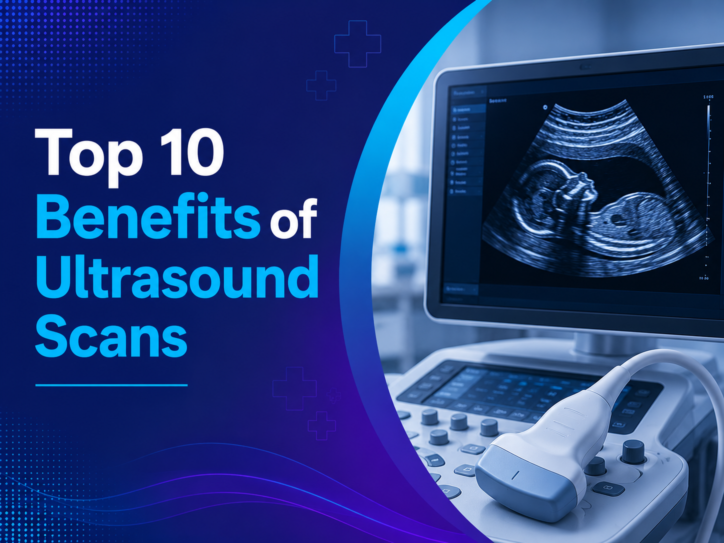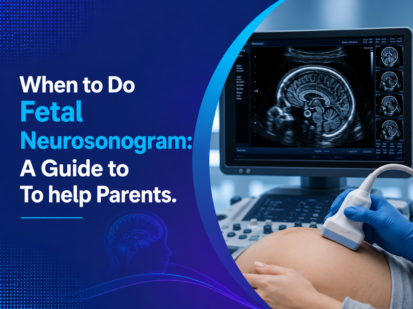
Prenatal care is about catching small concerns before they become big ones, and ultrasound is one of its most powerful tools. Ultrasounds During Pregnancy show how your baby is growing, where the placenta sits, and whether anything needs closer attention. This guide explains why these scans matter, what the World Health Organization recommends, and how to get the most from them.
Why Ultrasounds During Pregnancy Are Essential: The Short Answer
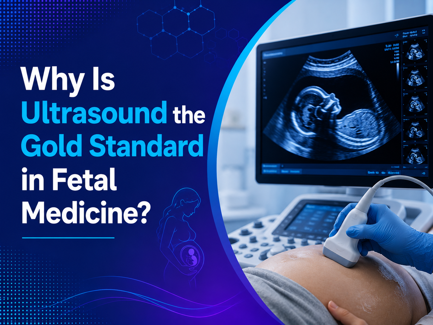
Ultrasound is essential in prenatal care because it gives doctors direct, safe, radiation-free information that examinations and blood tests cannot. It helps in six main ways:
- Confirms an accurate due date
- Finds twins or more early
- Checks the baby’s development
- Checks the placenta and amniotic fluid
- Monitors growth and well-being
- Helps plan a safer delivery
What the WHO Recommends About Ultrasounds During Pregnancy
The World Health Organization recommends at least one ultrasound scan before 24 weeks of pregnancy. Its purpose is to estimate gestational age, improve detection of fetal anomalies and multiple pregnancies, reduce unnecessary induction for post-term pregnancy, and improve the pregnancy experience. WHO reviewed the evidence again in 2021 and kept the recommendation.
WHO does not advise a routine scan after 24 weeks for women who already had an early one. Further scans are added when there is a medical reason, such as a complication or a high-risk condition.
1. Accurate Dating Protects Mother and Baby
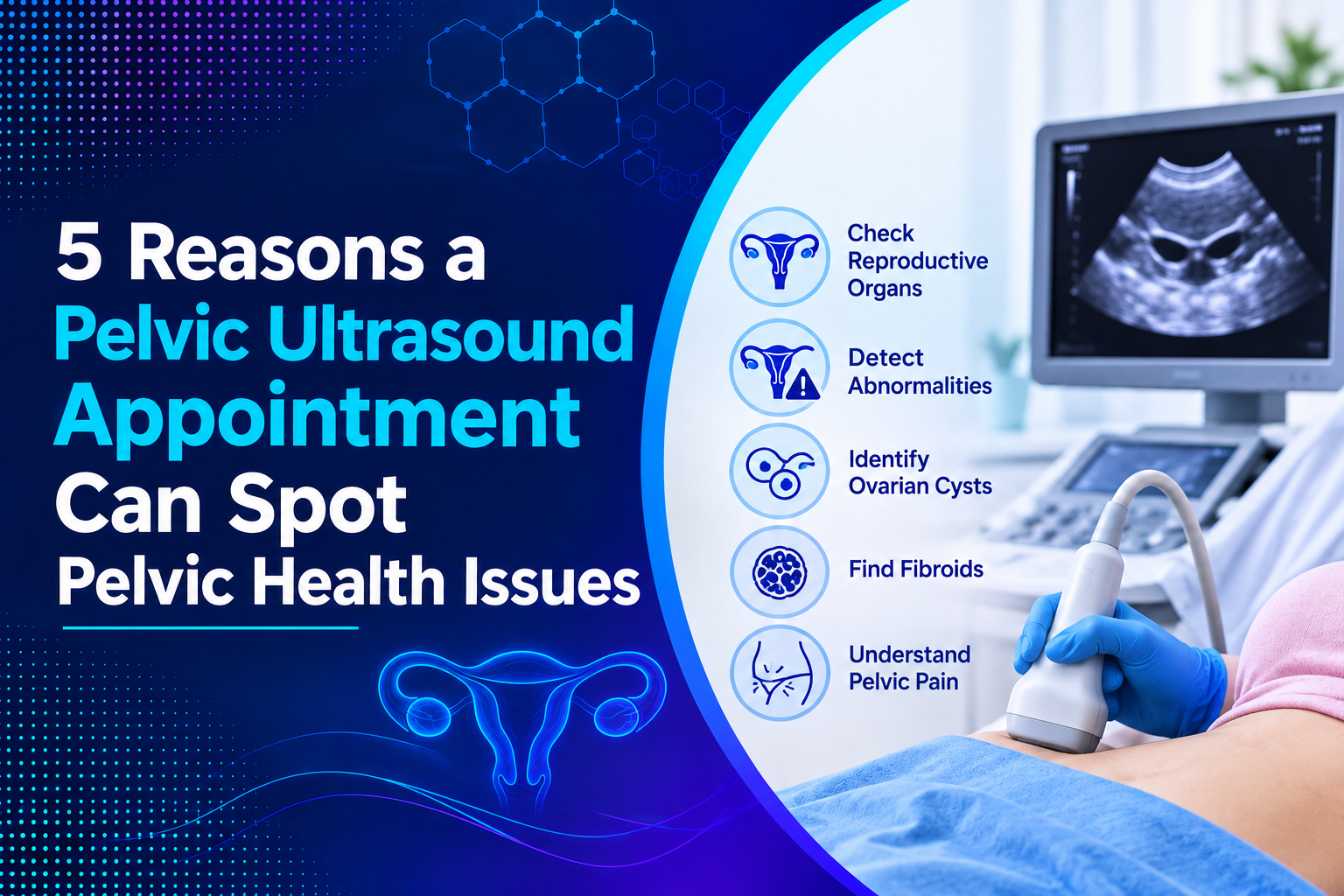
Knowing exactly how far along you are affects almost every decision in pregnancy. It guides the timing of screening tests, the reading of growth, and the plan for delivery. Dating by ultrasound is most accurate in the first trimester, which is why early scans matter. Women looking for a Pregnancy ultrasound in Indore for dating can book this scan once the pregnancy is confirmed.
2. Early Detection of Twins and Multiple Pregnancies
A twin pregnancy needs different monitoring from a single one. Ultrasound shows the number of babies and, in multiples, whether they share a placenta. This knowledge shapes how often you are scanned and how carefully growth is tracked.
3. Checking Your Baby’s Development and Anatomy
The detailed anatomy scan, usually at 18 to 22 weeks, examines the brain, heart, spine, face, abdomen, and limbs. Finding a concern early gives parents and doctors time to plan, arrange extra tests, or prepare for specialist care at birth.
4. Checking the Placenta and Amniotic Fluid
The placenta feeds your baby, and its position matters. If it lies low and covers the cervix, a condition called placenta previa, your doctor can plan safe monitoring and delivery. Ultrasound also measures amniotic fluid, since too much or too little can signal problems.
5. Monitoring Growth and Well-Being
Later scans track whether the baby is growing steadily. For pregnancies with diabetes or high blood pressure, Doppler scans and a biophysical profile may be added to check blood flow and well-being. These tools help doctors act in time if growth slows.
6. Supporting Safer Delivery Planning
Near term, ultrasound can confirm the baby’s position, estimate size, and check the placenta and fluid. These details help your doctor decide on the safest approach to birth. Accurate dating also reduces unnecessary early induction.
7. Reassurance and Emotional Connection
Beyond medicine, scans give parents a real connection with their baby. Seeing the heartbeat and movements reassures many women and helps partners feel involved. For many families, this is one of the most meaningful parts of prenatal care.
How Many Ultrasounds During Pregnancy Do You Need?
The number depends on your health and your pregnancy. Most healthy pregnancies have a few well-timed scans.
| Stage | Typical Timing | Main Purpose |
| Early dating scan | Up to 14 weeks | Heartbeat, dating, number of babies |
| Screening scan | 11 to 14 weeks | Nuchal translucency screening |
| Anatomy scan | 18 to 22 weeks | Detailed check of the baby and placenta |
| Growth scan | Third trimester, if advised | Growth, position, and fluid |
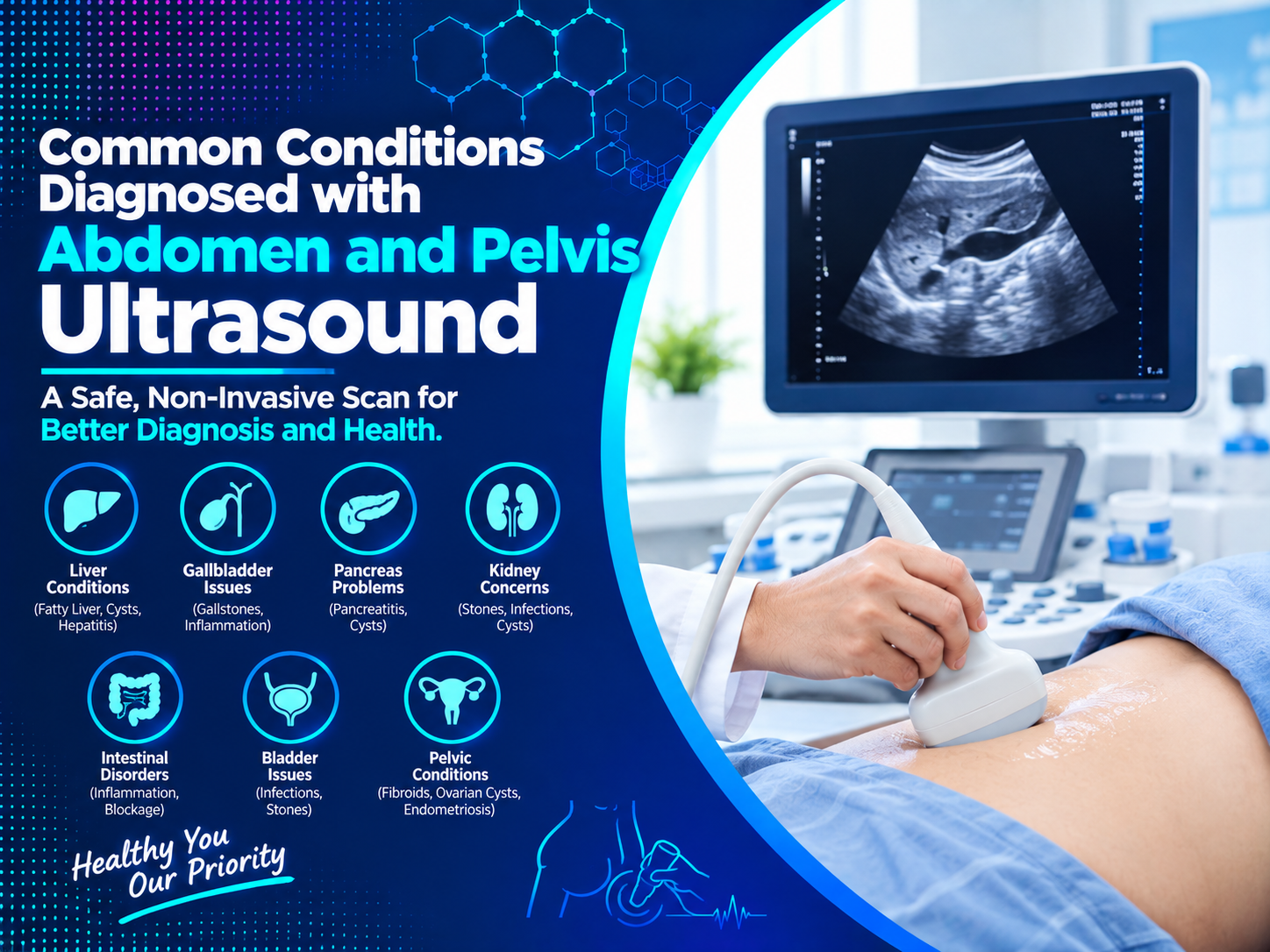
Families searching for ultrasound scans in Indore should ask their doctor which scans are needed for them, rather than booking extra ones without a reason.
Who May Need Extra Scans?
Healthy pregnancies often follow the standard schedule, but some situations call for closer watching.
- Diabetes or high blood pressure
- A twin or multiple pregnancy
- Bleeding or pain during pregnancy
- Reduced baby movements
- A baby measuring smaller or larger than expected
- A previous pregnancy complication
An extra scan in these cases is a sign of careful care, not a reason to worry.
Understanding Your USG Report
In India, scan reports are often labeled USG, short for ultrasonography. A typical report lists the baby’s heartbeat, estimated due date, fetal measurements, estimated weight, placenta position, and amniotic fluid. A clear USG in Indore report should explain each finding in simple terms, so you know what is normal and what needs follow-up.
Are Ultrasounds During Pregnancy Safe?
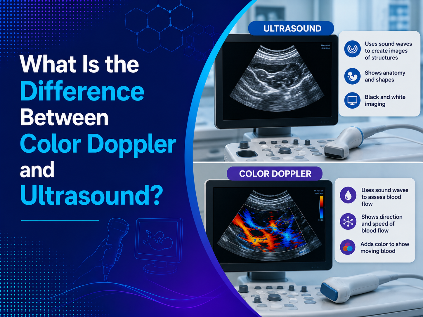
Ultrasound uses sound waves rather than ionizing radiation, and it has been used in pregnancy care for decades. The safe approach is to scan when there is a good reason and to use trained professionals. A well-performed Ultrasound Test in Indore follows this principle and focuses on useful information.
What to Expect at Your Scan
- You lie down comfortably, and gel is applied to your abdomen.
- The sonographer moves the probe and takes measurements.
- Early in pregnancy, a vaginal probe may be used for clearer views.
- You can often see the screen and hear the heartbeat.
- A doctor reviews the images and shares the report with your gynecologist.
Common Myths About Pregnancy Scans
- Myth: More scans always mean better care. Fact: Scans work best when timed for a purpose.
- Myth: A 3D or 4D scan replaces routine scans. Fact: It adds detail but does not replace standard checks.
- Myth: A normal scan guarantees a healthy baby. Fact: Scans are valuable but cannot detect every condition.
What Ultrasounds Can and Cannot Do
- They can confirm dating, number, position, and many structural features
- They cannot detect every condition or birth difference
- Image quality depends on the baby’s position and your body
- A normal scan is reassuring, but it is not a guarantee
Tips to Prepare for Your Scans
- Carry your earlier reports and prescriptions
- Ask about bladder preparation
- Wear comfortable clothing
- Write down your questions
- Use one Diagnostic Centre in Indore for all your scans so your records stay together
Frequently Asked Questions
1. How many ultrasounds are essential during pregnancy?
WHO recommends at least one before 24 weeks. Most women have a few more, depending on their health and their doctor’s advice.
2. Are ultrasounds during pregnancy safe for the baby?
Yes. Ultrasound uses sound waves instead of radiation, and it is done only when there is a good reason.
3. When is the best time for a pregnancy ultrasound?
An early scan helps with dating, and the anatomy scan at 18 to 22 weeks gives a detailed check. Your doctor will plan the rest.
4. Can an ultrasound detect every problem?
No. It finds many conditions, but it cannot detect everything, so doctors also use blood tests and clinical checks.
5. Do I need a scan in every trimester?
Not always. Healthy pregnancies often need only a few scans, while high-risk pregnancies may need more.
Conclusion
Ultrasounds During Pregnancy are essential because they turn uncertainty into clear, useful information at every stage. Used wisely and at the right time, they protect both mother and baby and support confident decisions.
Book your pregnancy scan at Ultrascan Diagnostic Centre today. Call ahead or walk in to plan your schedule. This article is for general information and does not replace advice from your gynecologist.