Seeing your baby’s face clearly for the first time is a moment many expecting parents describe as unforgettable. 3D and 4D Sonography has transformed this experience, moving beyond the flat, grainy outlines of traditional imaging to offer remarkably detailed, lifelike views of your baby before birth. This guide explains how this technology works, what makes it different from standard scans, and why it has become such a meaningful part of the pregnancy journey for so many families.
What Is 3D and 4D Sonography?
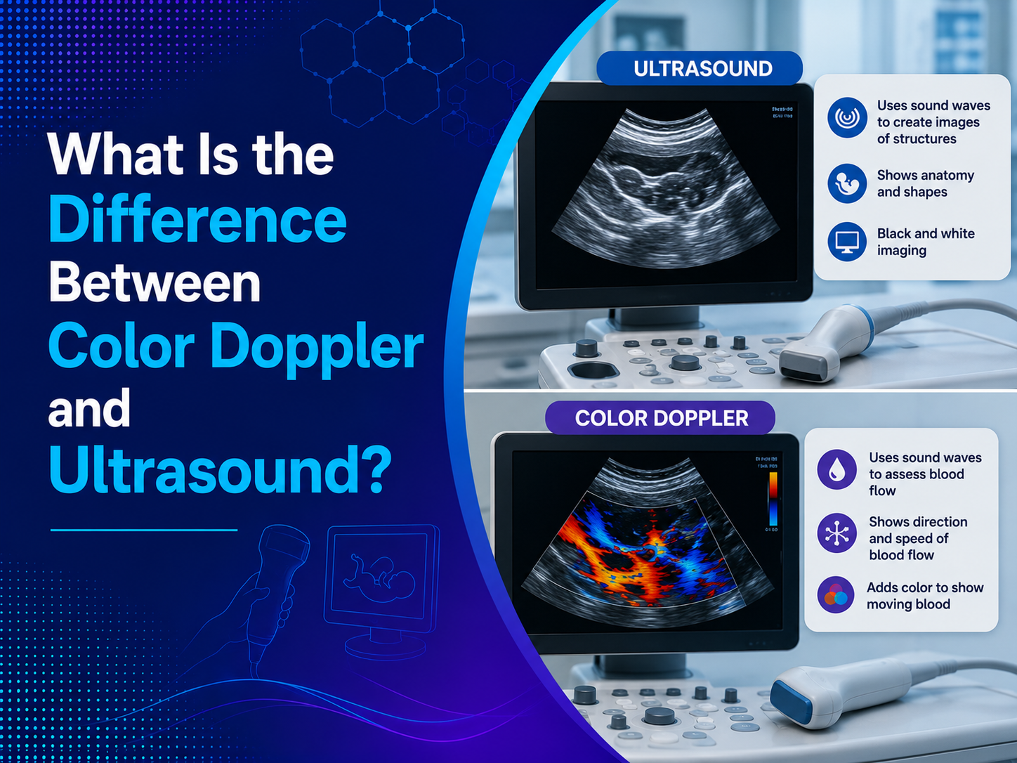
Traditional 2D ultrasound captures flat, cross-sectional images of the baby, which are highly useful for medical evaluation but can be difficult for parents to interpret visually. 3D and 4D Sonography builds on this same safe sound wave technology, but processes the data differently to create a three-dimensional image with depth and detail.
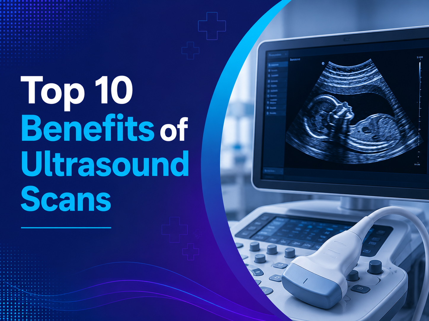
A 3D scan produces a still, sculpted image of the baby’s face and features, while 4D sonography adds the element of time, showing real-time movement such as yawning, stretching, or thumb-sucking. Both formats use the same non-invasive, radiation-free sound wave technology as a standard ultrasound, simply reconstructing the data into a more visually detailed format.
How 3D and 4D Sonography Differs From Standard Ultrasound
Understanding the distinction between these imaging types helps set realistic expectations for what each scan can show.
| Scan Type | Image Format | Best Used For |
| 2D Ultrasound | Flat, cross-sectional image | Routine medical evaluation, measurements, anatomy checks |
| 3D Sonography | Still, three-dimensional image | Detailed facial features and surface structures |
| 4D Sonography | Live, moving three-dimensional image | Observing real-time fetal movement and expressions |
While 2D remains the primary tool for medical assessment throughout pregnancy, 3D and 4D imaging adds a visually rich layer that many parents find deeply meaningful, without replacing the diagnostic value of standard scans.
The Emotional Value of 3D and 4D Fetal Imaging
Beyond the clinical benefits, there is a genuine emotional dimension to seeing your baby’s face and movements so clearly before birth. Many parents describe this moment as one of the first real steps toward bonding with their baby, transforming an abstract pregnancy into something tangible and deeply personal.
For partners and older siblings, a 3D and 4D Sonography session often becomes a shared family experience, offering a clear, relatable image rather than the harder-to-interpret outlines of a standard scan. This emotional connection can be especially meaningful for families going through fertility treatment or a pregnancy following previous loss.
Medical Uses of 3D and 4D Imaging Beyond Bonding
While parental bonding is a well-known benefit, 3D and 4D imaging also serves genuine medical purposes in certain situations.
- Detailed facial structure evaluation, useful when a cleft lip or palate is suspected
- Spine and limb assessment, offering a clearer view of skeletal structure in complex cases
- Enhanced visualization for suspected abnormalities, adding depth to findings from a standard scan
- Improved counseling discussions, helping doctors explain findings more clearly to parents
- Surgical and delivery planning, particularly in cases involving known structural concerns
In these situations, 3D and 4D imaging is used as a complementary tool alongside standard 2D ultrasound, rather than as a replacement for routine diagnostic scanning.
When Is the Best Time for a 3D or 4D Scan?
Timing plays an important role in getting the clearest possible images, since fetal position and available space in the womb change significantly throughout pregnancy.
Most doctors recommend scheduling a 3D or 4D scan between 26 and 32 weeks of pregnancy, when the baby has developed enough facial detail but still has sufficient amniotic fluid and room to move for clear imaging. Scanning too early may result in less defined features, while scanning too close to delivery can make clear facial views more difficult due to limited space and fetal positioning.
Is 3D and 4D Sonography Safe for the Baby?
Safety is one of the most common concerns parents raise before scheduling this type of scan, and the reassuring answer is that 3D and 4D sonography uses the exact same sound wave technology as standard 2D ultrasound. Neither format involves ionizing radiation, and both are considered safe when performed for appropriate medical or diagnostic purposes by trained professionals.
That said, health authorities generally recommend against excessive or purely recreational scanning without medical guidance, since ultrasound exposure should always serve a genuine purpose rather than being repeated unnecessarily. Scheduling your scan through a qualified medical facility, rather than an unregulated setting, ensures the procedure is performed appropriately and interpreted correctly.
What to Expect During Your 3D or 4D Sonography Session
Knowing what happens during the appointment can help you prepare and make the most of the experience.
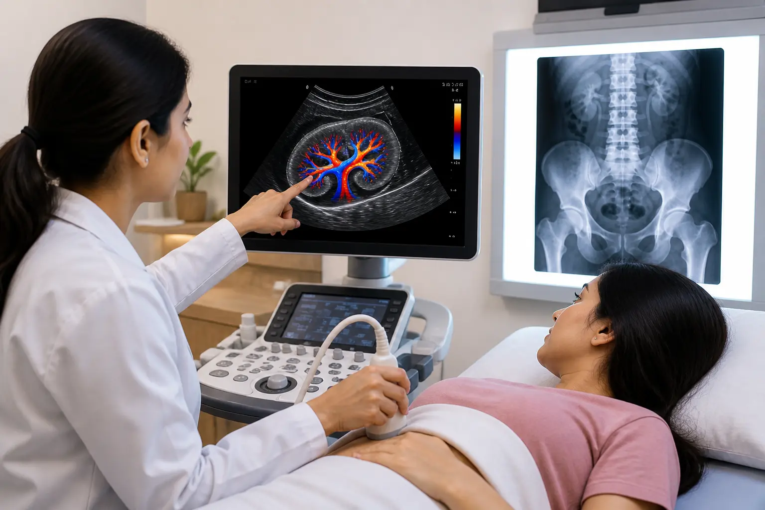
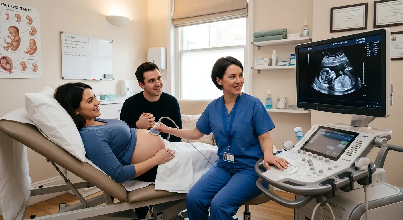
- You will lie down comfortably while a gel is applied to your abdomen.
- A sonographer moves the transducer gently over the area, searching for the clearest possible angle.
- The baby’s position and movement will influence how much detail is visible during the session.
- Images and, for 4D scans, short video clips are captured for you to keep.
- A radiologist reviews the results to confirm normal development alongside the detailed imagery.
Staying relaxed and well-hydrated before your appointment can sometimes help encourage fetal movement into a more favorable position for clearer images.
Choosing the Right Facility for Fetal Imaging
Since image clarity depends heavily on equipment quality and sonographer experience, choosing a well-equipped, medically supervised facility genuinely matters for both bonding and diagnostic purposes.
A Trusted Choice for Prenatal Imaging
Expecting parents searching for a reliable Pregnancy Sonography Centre in Indore typically look for a facility that combines advanced 3D and 4D technology with experienced radiologists who can confirm healthy development alongside the detailed imagery.
Comparing Options Across the City
When comparing different sonography centres in indore, it is worth asking specifically about the equipment used for 3D and 4D imaging, since not all facilities offer the same level of image clarity or detail.
Complete Prenatal Imaging Services
For patients researching sonography in Indore more broadly, choosing a centre that offers the full range of prenatal scans, from routine anatomy checks to detailed 3D and 4D imaging, simplifies coordinating care throughout pregnancy.
Specialized Cardiac Imaging When Needed
In cases where additional cardiac evaluation is recommended, access to Fetal Echocardiography in Indore at the same facility can make follow-up testing far more convenient for expecting parents.
Why Choose Ultrascan Diagnostic Centre
Ultrascan Diagnostic Centre is recognized as one of the best diagnostic centre in Indore for prenatal imaging, combining modern 3D and 4D equipment with experienced sonologists who prioritize both accuracy and a comfortable patient experience.
Consistent, Reliable Care Throughout Your Pregnancy
As a trusted Diagnostic Centre in Indore, Ultrascan supports expecting parents through every stage of pregnancy monitoring, from early confirmation scans to detailed imaging sessions later in the journey.
Frequently Asked Questions
1. Is 3D and 4D sonography safe for my baby?
Yes, both 3D and 4D sonography use the same safe sound wave technology as standard 2D ultrasound, with no ionizing radiation involved. Scans should still be performed by trained professionals for appropriate medical or diagnostic purposes rather than repeated excessively.
2. What is the best week of pregnancy to get a 3D or 4D scan?
Most doctors recommend scheduling between 26 and 32 weeks, when facial features are well developed and there is still enough amniotic fluid for clear imaging. Scanning outside this window may result in less detailed or clear images.
3. Can 3D and 4D imaging replace my routine anatomy scan?
No, 2D ultrasound remains the primary tool for medical evaluation and structural assessment throughout pregnancy. 3D and 4D imaging complements routine scans by adding visual detail, but does not replace standard diagnostic testing.
4. Why does my baby’s face sometimes look less clear during the scan?
Image clarity depends heavily on fetal position, amniotic fluid levels, and how the baby is facing the transducer at that moment. Babies who are facing away or positioned with limbs in front of the face can be more difficult to capture clearly.
5. Can 3D and 4D sonography detect birth defects?
While primarily used for detailed visualization, 3D and 4D imaging can sometimes help evaluate suspected facial or skeletal concerns identified during a standard scan. However, routine 2D ultrasound combined with other prenatal tests remains the primary tool for detecting structural abnormalities.
Conclusion
3D and 4D Sonography offers far more than a technical upgrade to traditional imaging, it creates a genuine emotional connection between parents and their baby well before delivery. While 2D ultrasound remains essential for medical evaluation, 3D and 4D imaging adds a meaningful, memorable layer to the pregnancy experience for countless families.
Book your 3D or 4D sonography session today at Ultrascan Diagnostic Centre and experience clear, detailed fetal imaging in a caring, professional setting. Walk in or call ahead to schedule your appointment and create a moment you will remember for years to come.