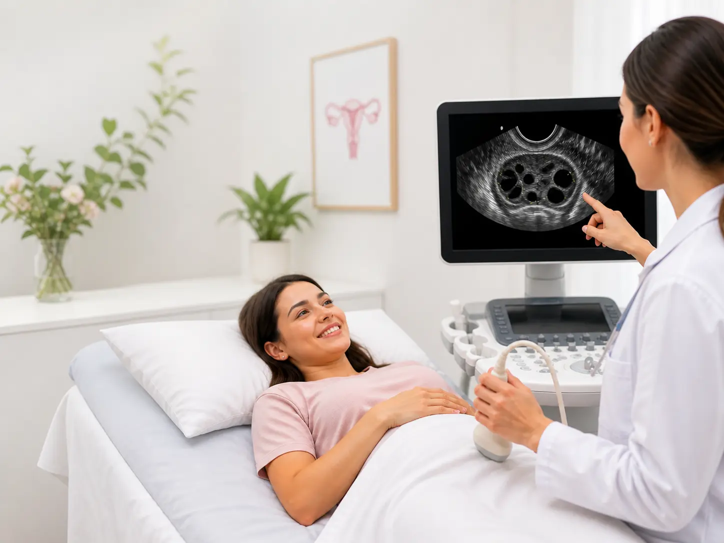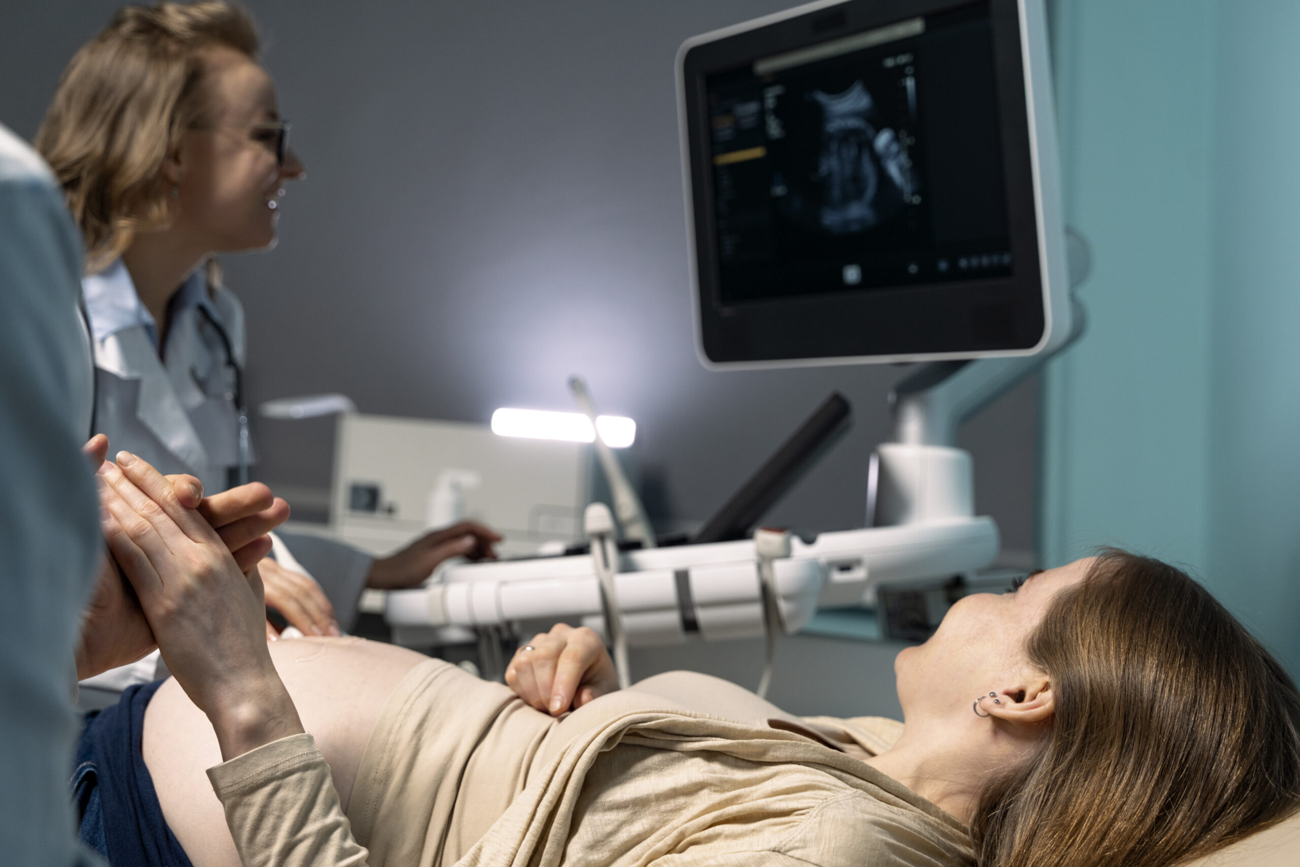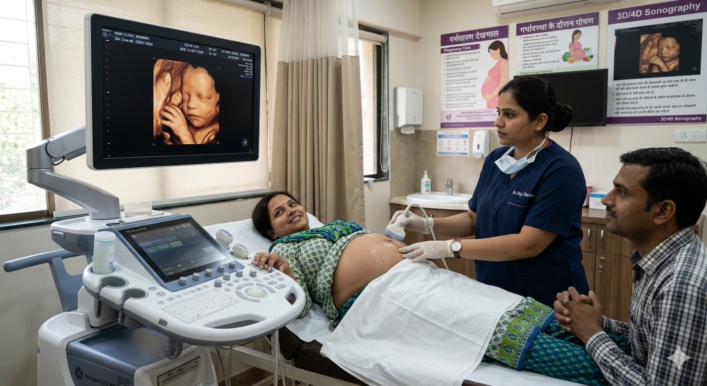
A fetal neurosonogram is a specialized prenatal ultrasound that provides detailed imaging of the developing fetal brain. Unlike routine obstetric scans, this targeted exam evaluates brain structures in depth — offering crucial insights into fetal neurological development.
Ultrascan Diagnostics offers multiple fetal neurosonogram-related searches in Indore, reflecting its position as the leading fetal neurosonogram clinic in the city.
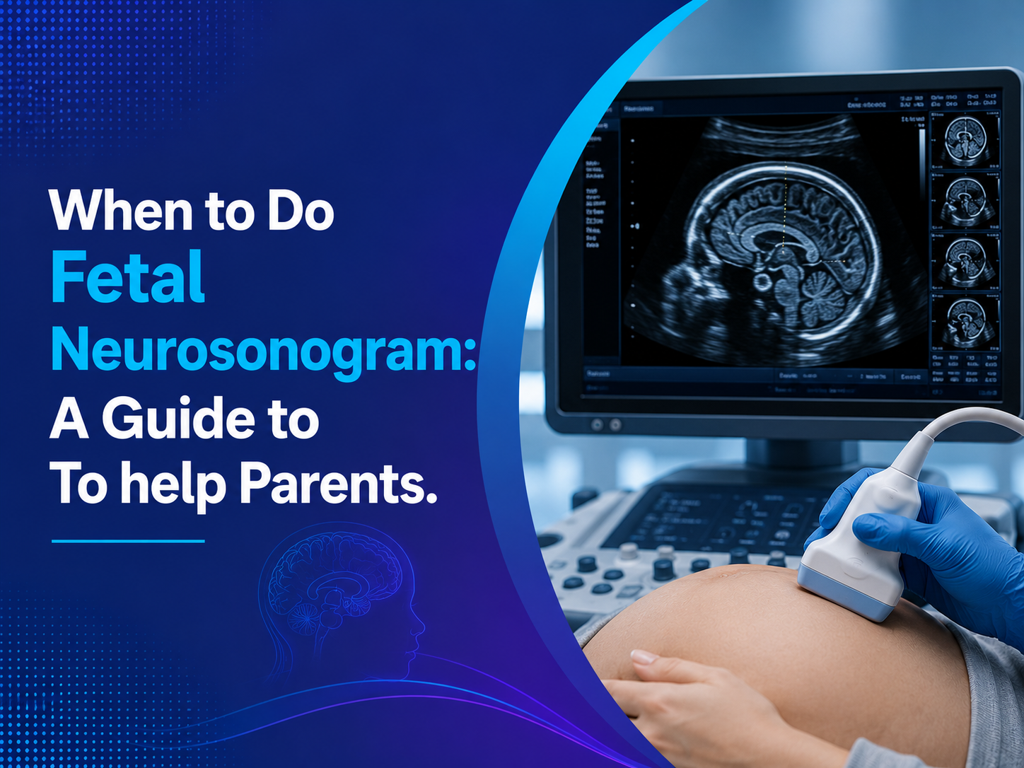
What Is a Fetal Neurosonogram?
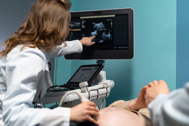
A fetal neurosonogram (also called a dedicated neurosonography or fetal brain scan) uses high-resolution ultrasound probes to image the fetal brain structures in three planes: axial, coronal, and sagittal. It is performed by a specialized radiologist with training in fetal medicine.
The scan evaluates:
- Cerebral cortex development and sulcation (brain folding)
- Corpus callosum — the structure connecting both brain halves
- Ventricles and cerebrospinal fluid spaces
- Posterior fossa (cerebellum and brainstem)
- Periventricular areas for haemorrhage or leukomalacia
- Brain midline structures
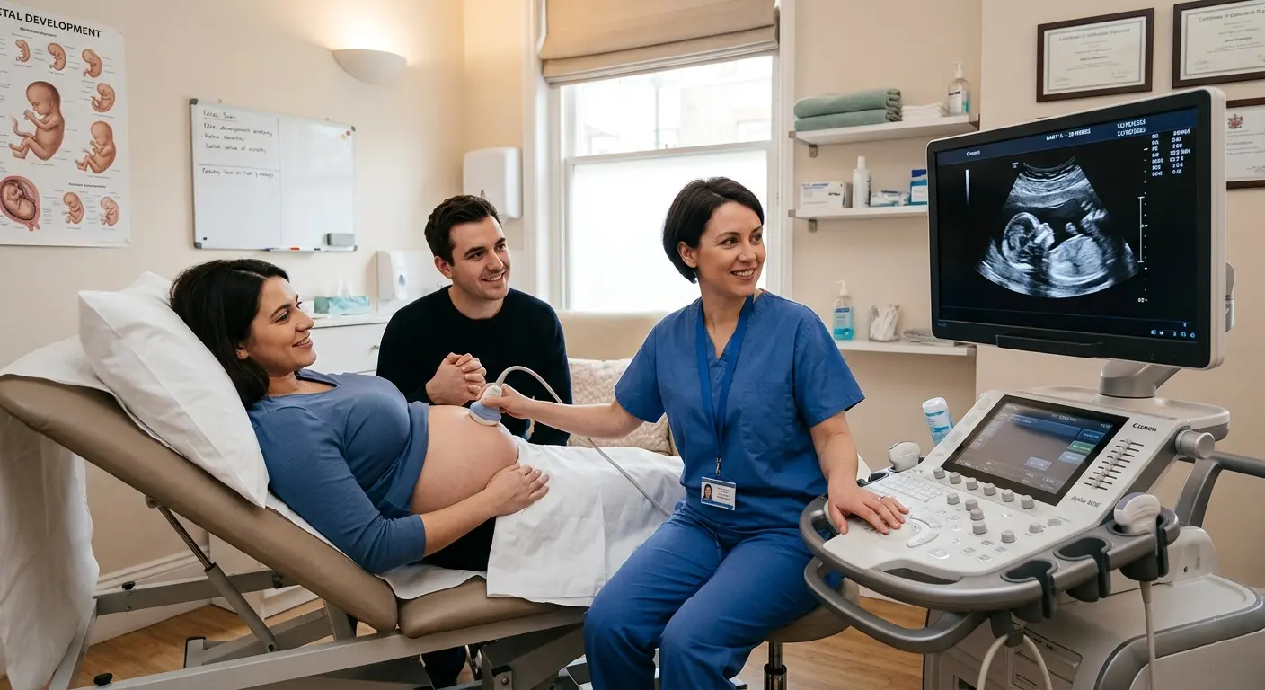
When Is a Fetal Neurosonogram Recommended?
Fetal neurosonogram is recommended in the following situations:
- Abnormal findings on routine anomaly scan (brain-related)
- Ventriculomegaly detected on standard anatomy scan
- Family history of neural tube defects or brain malformations
- Maternal infections (CMV, Toxoplasma, Zika) during pregnancy
- Exposure to teratogenic drugs in early pregnancy
- Advanced maternal age with increased chromosomal risk
- Previous child with neurological disorder
The best time to perform a fetal neurosonogram is between 28 and 32 weeks of gestation, when brain structures are sufficiently developed for detailed imaging.
What Can a Fetal Neurosonogram Detect?
This scan can help identify a range of fetal brain conditions, including:
- Ventriculomegaly (enlarged ventricles)
- Holoprosencephaly (failure of brain to divide into two hemispheres)
- Agenesis of the corpus callosum
- Dandy-Walker malformation
- Neural tube defects (e.g., spina bifida, anencephaly)
- Intracranial hemorrhage
- Microcephaly or macrocephaly
- Periventricular leukomalacia (PVL)
Early detection of these conditions allows families and clinicians to plan appropriate management, counseling, and delivery care.
Fetal Neurosonogram vs. Routine Anomaly Scan: What’s the Difference?
| Feature | Routine Anomaly Scan | Fetal Neurosonogram |
| Brain assessment | Basic (axial only) | Detailed (3 planes) |
| Corpus callosum | Not routinely evaluated | Specifically assessed |
| Duration | 30–45 min (full body) | 45–60 min (brain only) |
Frequently Asked Questions: Fetal Neurosonogram
Q1 : Is fetal neurosonogram safe for the baby?
Yes. Fetal neurosonogram uses diagnostic ultrasound with no ionizing radiation. It is considered safe at any gestational age and for repeated examinations.
Q2 : How should I prepare for a fetal neurosonogram?
No special preparation is required. You may be asked to drink water to fill the bladder if the scan is done in early pregnancy. Wear loose, comfortable clothing.
Q3: How long does the fetal neurosonogram take?
The scan typically takes 45 to 60 minutes. A detailed written report is usually provided on the same day.
Ultrascan Diagnostics: Indore’s #1 Fetal Neurosonogram Centre
- High-resolution ultrasound systems with multi-planar imaging capability
- Detailed written reports with images provided on same day
- Compassionate, patient-centred care for expecting parents
Final Words
Reviewed by senior radiologists at Ultrascan Diagnostics, Indore — recognized as the leading fetal neurosonogram clinic in Indore. Our team specializes in fetal medicine imaging, prenatal diagnosis, and high-risk pregnancy support.
Location: 451-G Greater Brajeshwari, Near Kerala Bakery, Pipliyahana Road, Indore | Contact: +91 78695 24599
Reach us at: ULTRASCAN-DIAGNOSTICS INDORE,MP