When your doctor recommends a kidney scan, the type of scan they choose makes a significant difference to what can be detected and how accurately a diagnosis can be made. Two of the most common kidney investigations are the KUB ultrasound scan and the renal Colour Doppler scan, and while they may sound similar, they reveal very different information about your kidney health.
Understanding what does a renal colour Doppler scan show – compared to what a standard KUB scan can detect – helps you appreciate why your doctor may have chosen one over the other, and what answers each test is designed to provide.
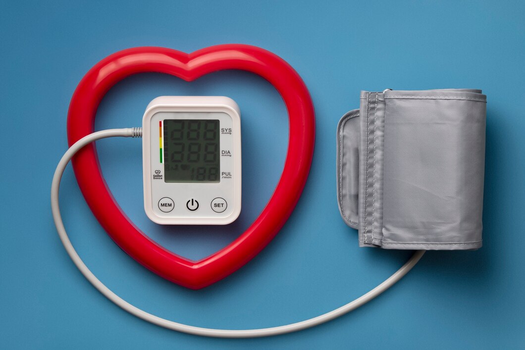
At Ultrascan Diagnostics, your trusted ultrasound diagnostic centre in Indore, we perform both KUB ultrasound scans and renal Colour Doppler scans using advanced imaging technology. This guide breaks down the differences in plain language so you can feel fully informed before your appointment.
What Is a KUB Ultrasound Scan and What Does It Detect?
KUB stands for Kidneys, Ureters, and Bladder. A KUB ultrasound scan uses standard greyscale (black-and-white) sound wave imaging to produce still and real-time images of these three organs. It is a structural scan – meaning it shows the shape, size, position, and texture of the kidneys, ureters, and bladder.
A KUB ultrasound scan is excellent at detecting:
• Kidney stones – their size and location within the kidney or ureter
• Hydronephrosis – swelling of the kidney due to a blockage preventing urine from draining
• Kidney cysts – fluid-filled sacs within or on the kidney
• Kidney tumours or masses – abnormal growths that need further investigation
• Changes in kidney size – a shrunken or enlarged kidney
• Bladder stones or bladder wall thickening
• Residual urine – how much urine remains in the bladder after urination
• Structural abnormalities of the urinary tract
A KUB ultrasound is the go-to scan for straightforward structural assessment. It is fast, non-invasive, affordable, and widely used as the first-line investigation for most urinary tract complaints – including pain in the flank, blood in urine, urinary tract infections, and difficulty passing urine.
What Does a Renal Colour Doppler Scan Show Beyond Structure?
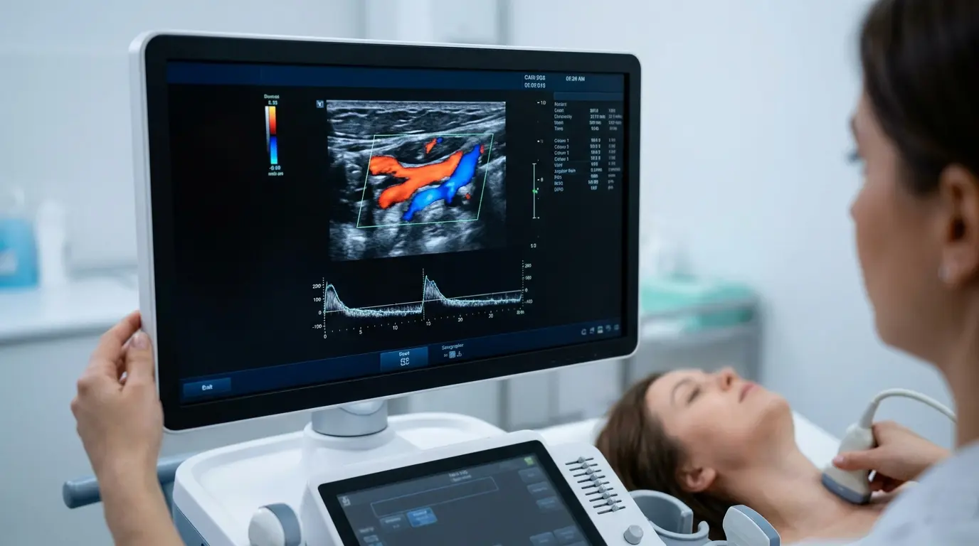
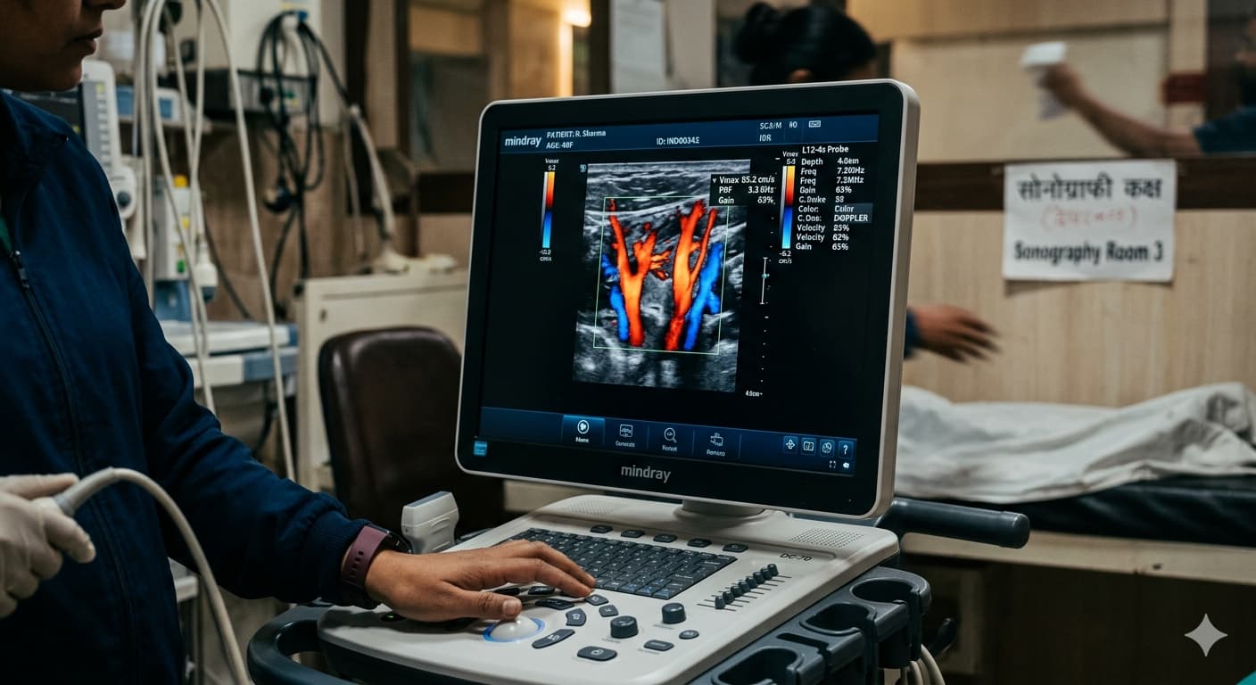
A renal Colour Doppler scan combines the structural imaging of a standard KUB ultrasound with an additional layer of information: blood flow. It uses the Doppler effect – the same principle that allows radar to detect moving objects – to track how blood moves through the arteries and veins supplying and draining the kidneys.
The scan produces colour-coded images where red and blue shades represent blood flowing toward and away from the probe, respectively. The brightness of the colour indicates flow speed. This real-time blood flow mapping reveals things that no structural scan – including a KUB – can show.
So what does a renal colour Doppler scan show that a KUB cannot? Here is the detailed answer:
1. Renal Artery Stenosis – Narrowing of the Kidney Arteries
Renal artery stenosis is a narrowing of one or both arteries supplying blood to the kidneys. It is one of the most common and frequently missed causes of high blood pressure that does not respond well to standard medication. A KUB scan cannot detect this at all, because it only shows structure.
A renal Colour Doppler scan, on the other hand, directly measures blood flow velocity through the renal arteries. Elevated peak systolic velocity and a raised resistive index are hallmark findings that point toward significant arterial narrowing. This finding can completely change the management plan – from adjusting medication to considering a procedure to open the narrowed artery.
2. Renal Vein Thrombosis – Blood Clots in the Kidney Veins
Renal vein thrombosis – a blood clot forming inside the vein draining a kidney – is a serious condition that can cause sudden flank pain, blood in urine, and in severe cases, acute kidney injury. It is invisible on a KUB ultrasound scan, which shows no blood flow information whatsoever.
A renal Colour Doppler scan detects this by showing absent or significantly reduced venous flow on the affected side – a direct indication that the vein is blocked. This is a critical diagnosis that requires prompt medical treatment, and Colour Doppler is the most accessible and reliable way to make it without exposing the patient to radiation or contrast dyes.
3. Resistive Index – A Window Into Kidney Function
One of the most clinically valuable parameters measured during a renal Colour Doppler scan is the resistive index (RI). This is a calculated value derived from blood flow measurements within the small arteries inside the kidney (interlobar or arcuate arteries). It reflects how much resistance the blood faces as it flows through kidney tissue.
An elevated resistive index – above 0.70 – suggests increased vascular resistance within the kidney, which can be associated with:
• Acute kidney injury (AKI)
• Chronic kidney disease (CKD) – severity correlates with RI level
• Obstructive nephropathy (blockage causing back-pressure damage)
• Rejection of a transplanted kidney
• Interstitial nephritis or other inflammatory conditions
A KUB ultrasound scan gives no information about resistive index whatsoever. This parameter is entirely unique to Colour Doppler assessment and gives clinicians vital functional insight into how well the kidneys are perfused – even before kidney function blood tests (creatinine, eGFR) show significant changes.
4. Kidney Transplant Monitoring
For patients who have received a kidney transplant, regular Colour Doppler scans are a standard and essential part of follow-up care. The transplanted kidney sits in an unusual position (typically in the pelvis rather than the back), and its blood supply comes from surgically created connections to nearby vessels. These need to be monitored carefully for complications including:
• Renal artery stenosis at the anastomosis site (where arteries were joined)
• Renal vein thrombosis
• Transplant rejection – signalled by rising resistive index
• Arteriovenous fistula – an abnormal connection between an artery and vein
• Perinephric collections (fluid around the transplant kidney)
None of these blood-flow-related complications can be assessed with a KUB scan. Colour Doppler is irreplaceable in this context, and any centre offering transplant monitoring must be equipped to perform it reliably.
5. Differentiation of Kidney Masses – Benign vs Potentially Malignant
When a KUB scan identifies a mass or lesion within the kidney, the next step is often to characterise it further – to determine whether it is likely to be benign (such as an angiomyolipoma or a simple cyst) or potentially malignant (such as a renal cell carcinoma).
A renal Colour Doppler scan contributes by assessing the vascularity of the mass – whether it has its own blood supply. Hypervascular masses (those with significant internal blood flow) are more likely to be malignant. This additional information helps guide the decision on whether a biopsy, further imaging (CT or MRI), or close monitoring is the right next step.
KUB Ultrasound vs Renal Colour Doppler Scan: A Quick Comparison
Here is a side-by-side summary to make the difference clear:
• Kidney size and shape → KUB: Yes | Renal Colour Doppler: Yes
• Kidney stones → KUB: Yes | Renal Colour Doppler: Yes
• Hydronephrosis (kidney swelling) → KUB: Yes | Renal Colour Doppler: Yes
• Kidney cysts and masses → KUB: Yes | Renal Colour Doppler: Yes (plus vascularity)
• Renal artery stenosis → KUB: No | Renal Colour Doppler: Yes
• Renal vein thrombosis → KUB: No | Renal Colour Doppler: Yes
• Resistive index / kidney perfusion → KUB: No | Renal Colour Doppler: Yes
• Transplant kidney monitoring → KUB: Partial | Renal Colour Doppler: Comprehensive
• Mass vascularity (benign vs malignant clues) → KUB: No | Renal Colour Doppler: Yes
• Blood flow velocity measurements → KUB: No | Renal Colour Doppler: Yes
The takeaway is clear: a KUB scan is your starting point for structural kidney assessment. A renal Colour Doppler scan is the next level – offering everything a KUB shows, plus critical blood flow information that structural imaging alone simply cannot provide.
When Would Your Doctor Recommend a Renal Colour Doppler Over a KUB Scan?
Your doctor is likely to choose a renal Colour Doppler scan over – or in addition to – a KUB ultrasound if you present with any of the following:
• High blood pressure that is difficult to control with standard medications
• Suspected renal artery stenosis or renal vascular disease
• Sudden unexplained deterioration in kidney function
• Blood in urine combined with concern about a kidney mass
• History of a kidney transplant requiring regular monitoring
• Chronic kidney disease with a need to track progression through resistive index
• Suspected blood clot in a renal vein
• Post-procedure assessment after a kidney-related intervention
• Unexplained flank pain alongside abnormal kidney function blood tests
What to Expect During a Renal Colour Doppler Scan at Ultrascan Diagnostics
If your doctor has referred you for a renal Colour Doppler scan at our ultrasound scan in Indore centre, here is what the process involves:
• Preparation: You will typically be asked to fast for 4 to 6 hours before the scan to reduce bowel gas that can obscure abdominal vessels. Staying well-hydrated is recommended. Confirm any specific instructions at the time of booking.
• Positioning: You will lie on your back and possibly on your side during the scan. The probe is gently moved across your abdomen and flanks.
• Duration: A renal Colour Doppler scan typically takes 30 to 45 minutes, slightly longer than a standard KUB scan because multiple blood flow measurements are taken from each kidney.
• Sensation: The scan is painless. A warm, water-based gel is applied to the skin, and you may feel gentle pressure from the probe.
• Results: A detailed written report is generated promptly and shared with your referring doctor. Our radiologists are available to discuss findings where needed.
Ultrascan Diagnostics is one of the nearest ultrasound centres in the Pipliyahana area of Indore, offering high-resolution ultrasound services for both renal Colour Doppler and KUB scans, with same-day reporting and a patient-first approach at every step.
Frequently Asked Questions About Renal Colour Doppler vs KUB Scans
Q1. What does a renal colour Doppler scan show that blood tests cannot?
Blood tests such as serum creatinine and eGFR (estimated glomerular filtration rate) tell you how well the kidneys are filtering blood overall – but they cannot tell you why kidney function is reduced, which kidney is affected, or what is happening in the blood vessels. A renal Colour Doppler scan shows blood flow velocity, the resistive index, and vascular anatomy directly. This means it can identify renal artery stenosis, renal vein thrombosis, and vascular resistance patterns long before blood tests show significant abnormality – making it a powerful early diagnostic tool. The combination of both investigations provides the fullest picture of kidney health.
Q2. Can a KUB scan detect high blood pressure caused by a kidney artery problem?
No – a standard KUB ultrasound scan cannot detect renovascular hypertension (high blood pressure caused by narrowing of the renal arteries). A KUB scan shows only the structure of the kidney – its size, shape, and the presence of stones or cysts. It provides no information about blood flow velocity, arterial narrowing, or vascular resistance. A renal Colour Doppler scan is specifically required to evaluate the renal arteries and identify stenosis. If you have high blood pressure that does not respond normally to medication, your doctor may request a renal Colour Doppler to rule out a vascular cause before exploring other explanations.
Q3. How often should kidney transplant patients have a renal Colour Doppler scan?
The frequency of post-transplant renal Colour Doppler scans depends on the clinical protocol of your transplant team and how well the transplanted kidney is functioning. In the immediate post-operative period (first few days to weeks), scans may be performed daily or every few days to ensure the anastomotic vessels are open and blood flow is stable. Once the transplant stabilises, scans are typically done at regular intervals – often at 1 month, 3 months, 6 months, and 12 months post-transplant, and then annually. Any episode of rising creatinine, decreased urine output, or pain over the transplant site warrants an immediate Colour Doppler assessment. Your transplant nephrologist will provide a personalised monitoring schedule. At Ultrascan Diagnostics, we accommodate urgent and scheduled renal Colour Doppler appointments for transplant patients with priority.