In the realm of prenatal care, few technological advancements have made as significant an impact as colour Doppler sonography. This powerful imaging technique has revolutionized the way healthcare professionals detect and manage congenital heart defects, offering hope and improved outcomes for countless families. In this article, we’ll explore the crucial role of colour Doppler sonography in identifying these life-altering conditions and the importance of early detection.
Understanding Congenital Heart Defects
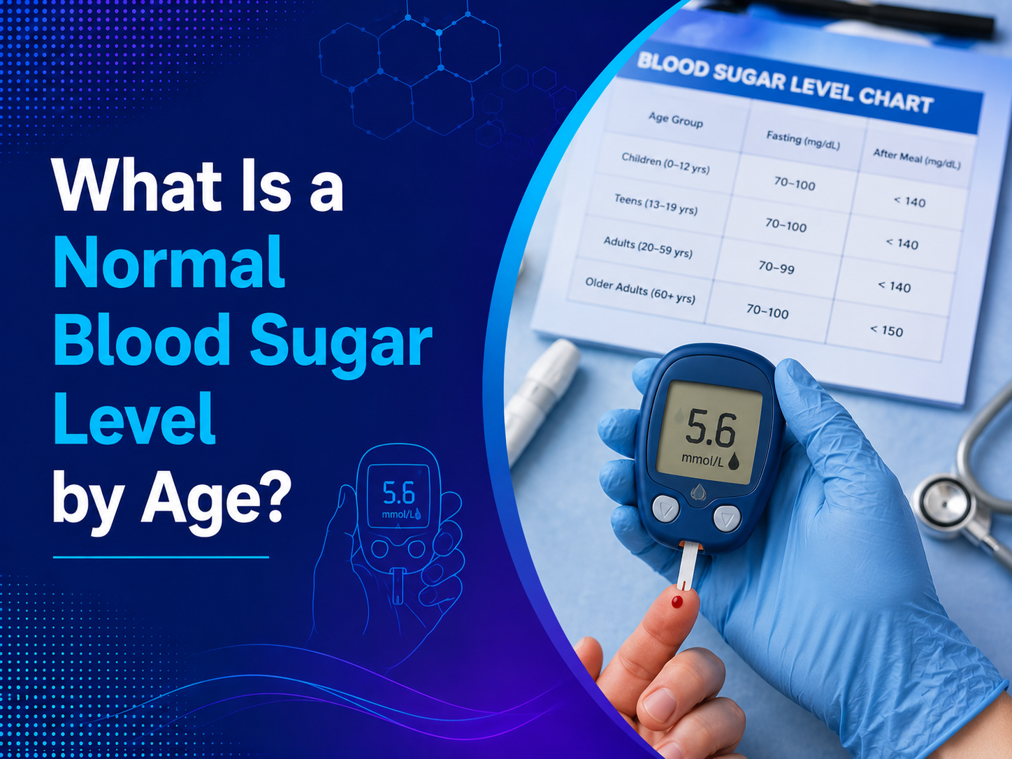
Congenital heart defects (CHDs) are structural problems with the heart that are present at birth. These defects can range from simple issues that resolve on their own to complex conditions requiring immediate intervention. CHDs affect nearly 1% of all births worldwide, making them the most common type of birth defect.
Types of Congenital Heart Defects
- Septal Defects: Holes in the wall separating heart chambers
- Valve Defects: Abnormalities in heart valves
- Outflow Tract Obstruction: Narrowing of major blood vessels
- Complex Defects: Combination of multiple heart abnormalities
The Role of Colour Doppler Sonography
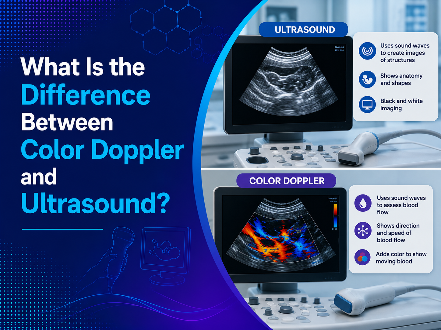
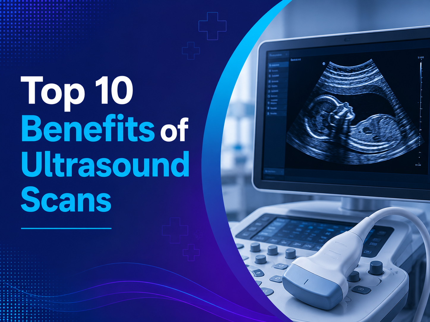
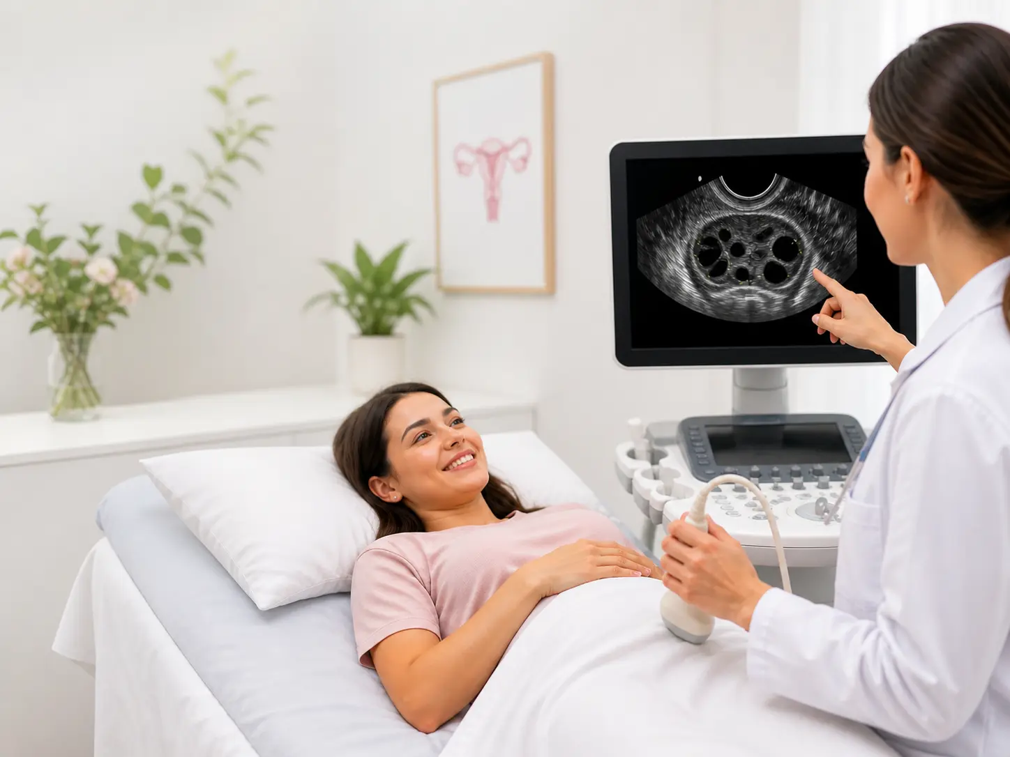
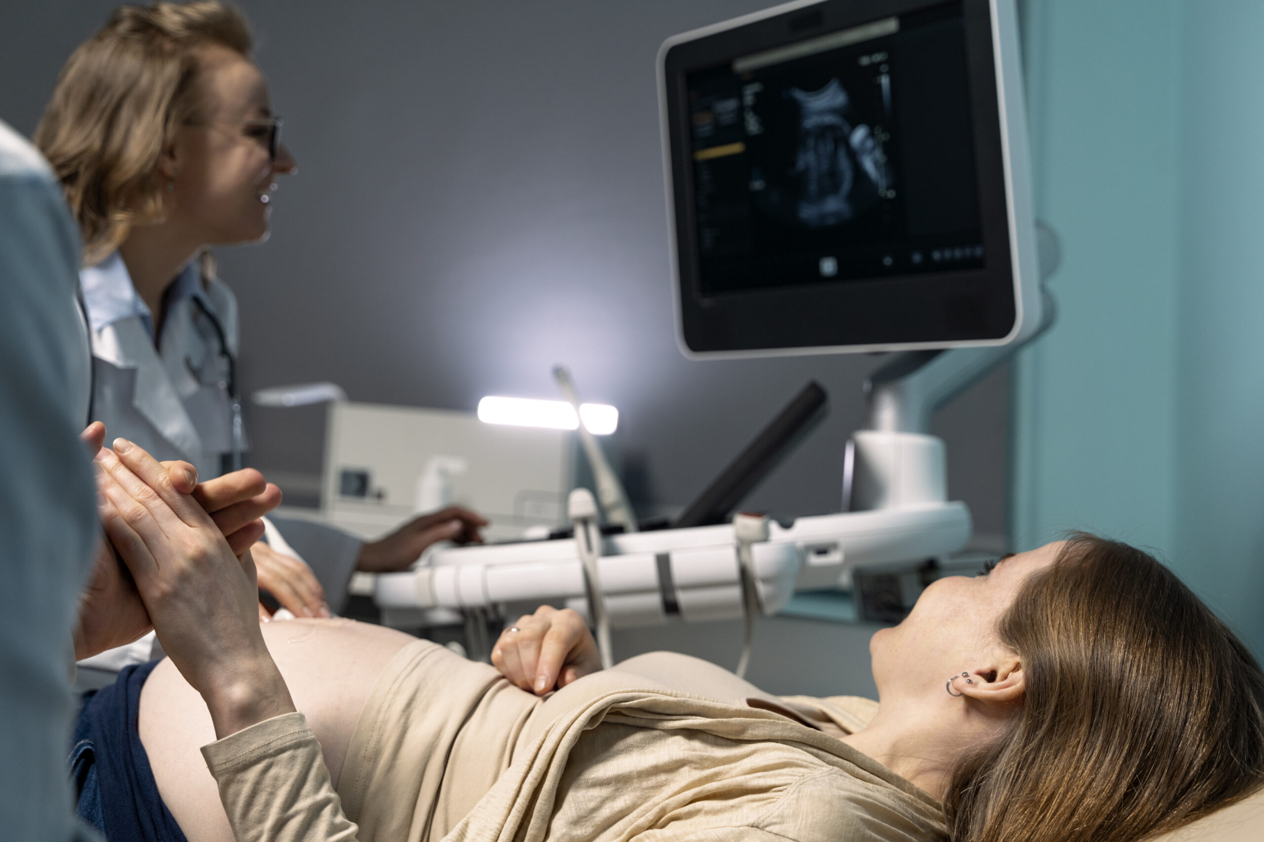
Colour Doppler sonography is an advanced ultrasound technique that uses sound waves to create detailed images of blood flow within the heart and blood vessels. This non-invasive procedure has become an indispensable tool in the detection and assessment of CHDs during pregnancy.
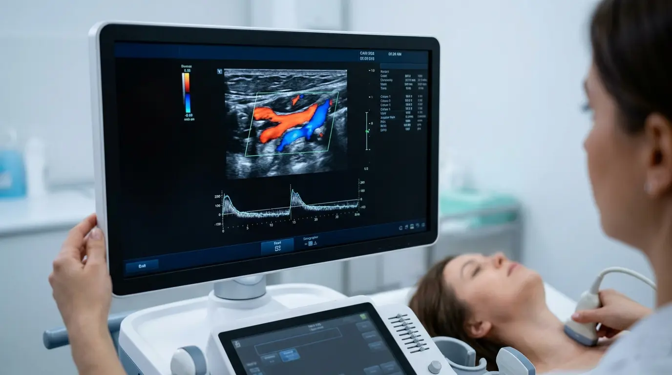
How Colour Doppler Works
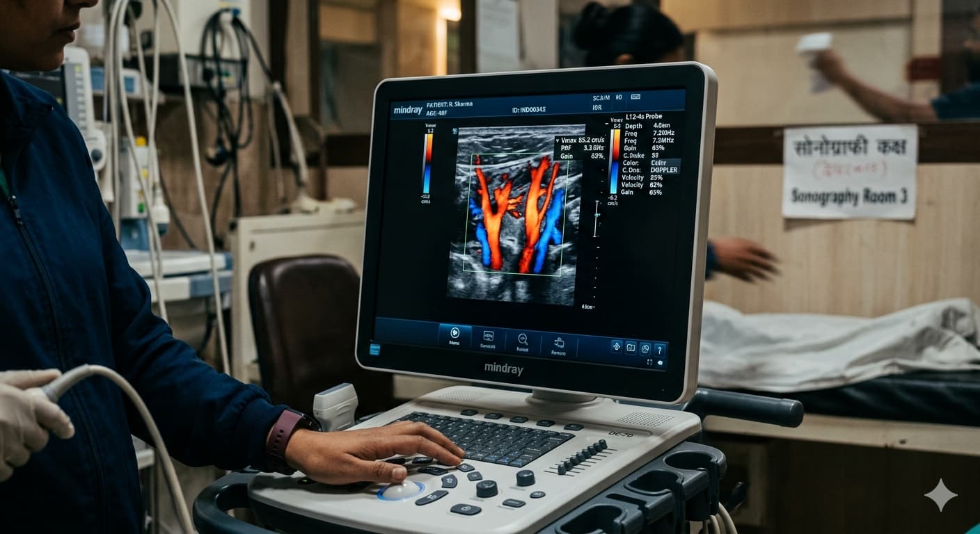
The technology behind colour Doppler sonography is based on the Doppler effect, which measures the change in frequency of sound waves as they bounce off moving objects, such as blood cells. This information is then translated into color-coded images that represent the direction and speed of blood flow. Red typically indicates blood flowing towards the transducer, while blue represents blood flowing away from it.
The Importance of Early Detection
Identifying CHDs as early as possible during pregnancy is crucial for several reasons:
- Improved Planning: Early detection allows healthcare providers and parents to plan for appropriate care immediately after birth.
- Better Outcomes: Some CHDs may require intervention shortly after birth. Early detection ensures that the necessary medical team and resources are available.
- Emotional Preparation: Knowing about a CHD in advance gives parents time to educate themselves and prepare emotionally for the challenges ahead.
- Potential for Fetal Intervention: In some cases, certain heart defects can be treated while the baby is still in the womb.
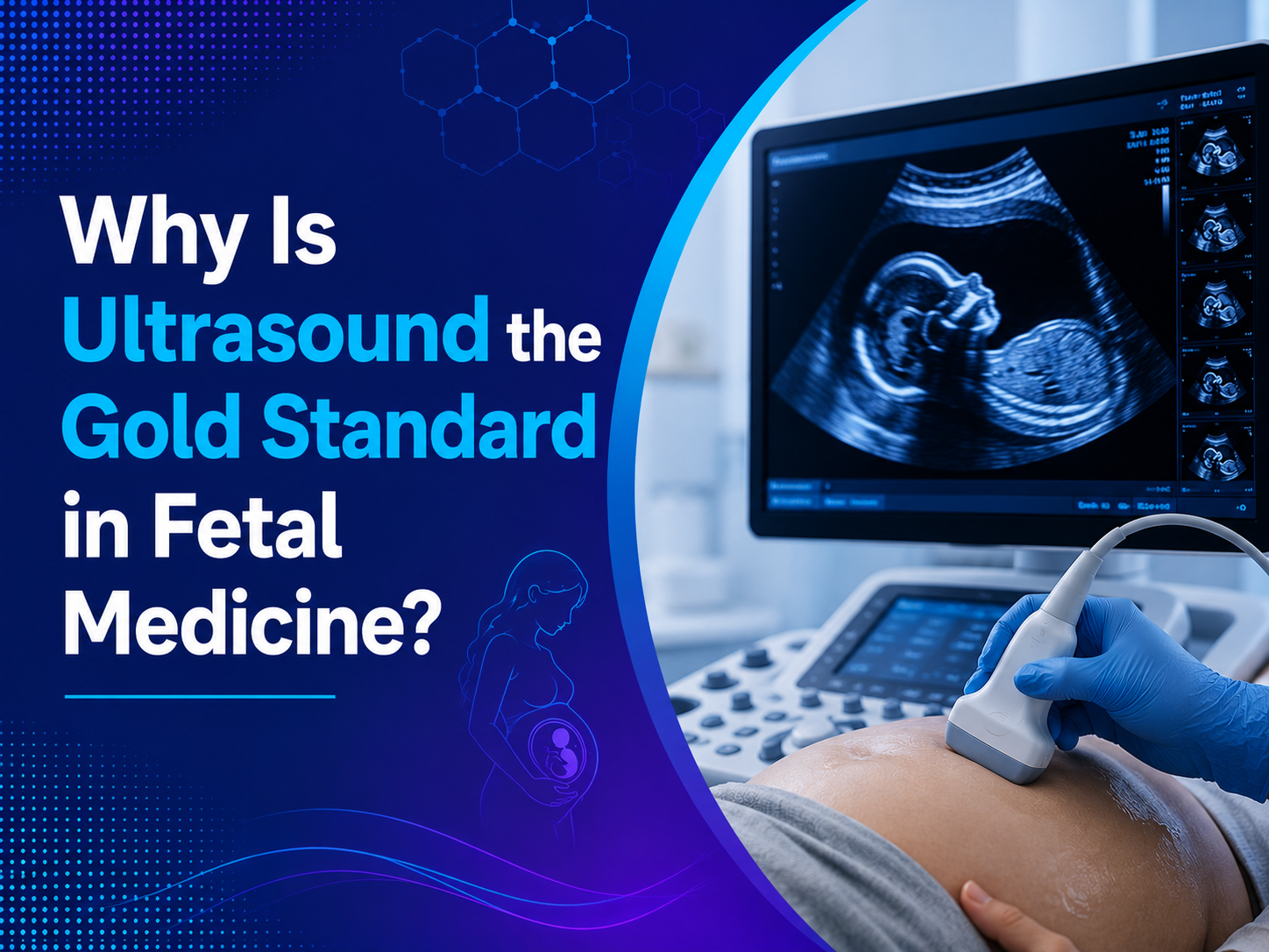
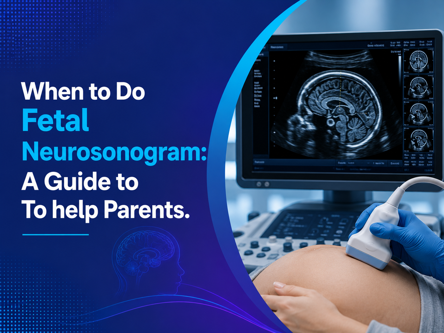
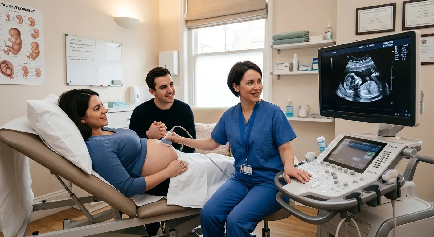
The Process of Fetal Heart Screening
Fetal heart screening typically occurs during the routine anatomy scan, which is usually performed between 18 and 22 weeks of pregnancy. However, some heart defects can be detected as early as 12 weeks gestation.
Steps in Fetal Heart Screening
- Four-Chamber View: This basic view assesses the size, position, and structure of the heart’s four chambers.
- Outflow Tracts: Examination of the major blood vessels leaving the heart.
- Three-Vessel View: Assessment of the relationship between the pulmonary artery, aorta, and superior vena cava.
- Colour Doppler Assessment: Evaluation of blood flow patterns within the heart and major vessels.
Advanced Fetal Echocardiography
In cases where a CHD is suspected or for high-risk pregnancies, a detailed fetal echocardiogram may be recommended. This specialized ultrasound examination provides a comprehensive assessment of the fetal heart structure and function.
Benefits of Fetal Echocardiography
- Detailed Imaging: High-resolution images of the fetal heart and blood vessels.
- Functional Assessment: Evaluation of heart rhythm and contractility.
- Precise Diagnosis: Accurate identification of specific heart defects.
- Treatment Planning: Allows for development of a tailored treatment plan before birth.
The Importance of Choosing the Right Imaging Center
When it comes to prenatal cardiac imaging, the quality of the equipment and the expertise of the sonographer are paramount. That’s why selecting the best sonography centre in Indore is crucial for expectant parents.
What to Look for in a Sonography Center in Indore
- State-of-the-Art Equipment: Advanced ultrasound machines with colour Doppler capabilities are essential for accurate fetal heart imaging.
- Experienced Sonographers: Skilled technicians with specialized training in fetal cardiac imaging can make a significant difference in detecting subtle abnormalities.
- Comprehensive Services: Look for centers that offer a full range of prenatal imaging services, including detailed fetal echocardiography.
- Collaborative Approach: The best centers work closely with maternal-fetal medicine specialists and pediatric cardiologists to ensure comprehensive care.
- Comfortable Environment: A welcoming atmosphere can help reduce anxiety during these important examinations.
Sonography in Indore: A Growing Field
The field of sonography in Indore has seen significant advancements in recent years, with an increasing number of specialized centers offering cutting-edge prenatal imaging services. This growth has made it easier for expectant parents to access high-quality fetal cardiac screening close to home.
Advancements in Sonography Indore
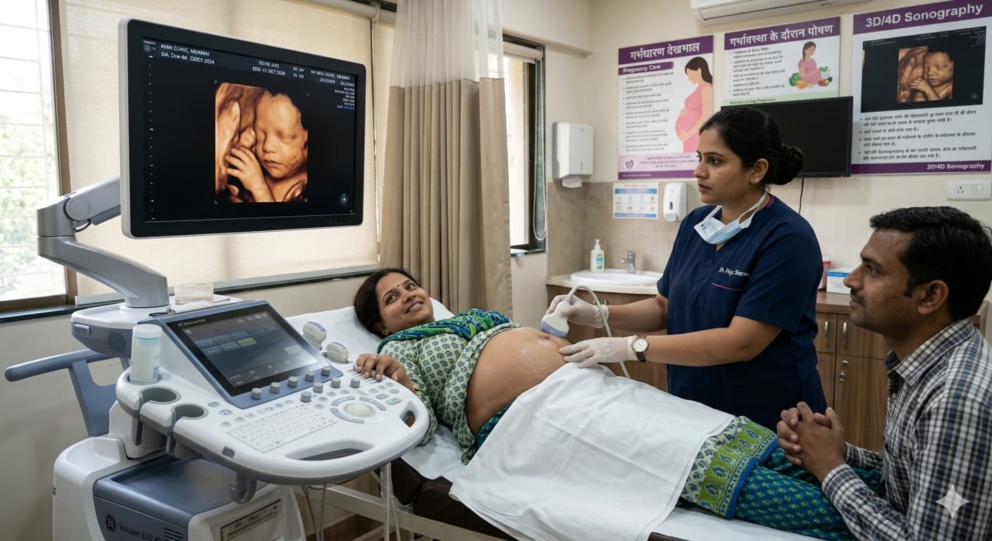
- 3D and 4D Imaging: Many centers now offer 3D and 4D ultrasound services, providing detailed views of the fetal heart and other structures.
- Telemedicine Integration: Some facilities offer remote consultations with pediatric cardiologists, expanding access to expert care.
- AI-Assisted Interpretation: Emerging artificial intelligence technologies are being developed to assist in analyzing fetal heart images, potentially improving accuracy and consistency in diagnosis.
The Role of Renal Doppler Test in Indore
While our focus is on fetal cardiac imaging, it’s worth noting that colour Doppler technology has applications beyond heart assessment. The renal Doppler test in Indore is an important diagnostic tool for evaluating kidney function in both pregnant women and the general population.
Benefits of Renal Doppler Testing
- Non-invasive: The test is painless and does not require any injections or contrast agents.
- Quick and Efficient: Results are available immediately, allowing for prompt decision-making.
- Comprehensive: It provides detailed information about blood flow in the kidneys, helping diagnose conditions such as renal artery stenosis or kidney transplant rejection.
The Cost of Advanced Fetal Imaging
While the benefits of advanced fetal imaging techniques are clear, many expectant parents are concerned about the associated costs. In Indore, the prices for specialized ultrasound services can vary depending on the facility and the complexity of the examination.
3D Sonography in Pregnancy Price
The cost of 3D sonography in pregnancy typically ranges from ₹1,500 to ₹3,000, depending on the center and the specific package chosen. This price usually includes a set of printed images and, in some cases, a digital copy of the scan.
4D Sonography in Pregnancy Price
4D sonography, which provides moving 3D images in real-time, is generally more expensive. Prices for 4D scans in Indore typically range from ₹3,000 to ₹6,000. Many centers offer package deals that include multiple scans throughout the pregnancy at a discounted rate.
It’s important to note that while 3D and 4D scans can provide beautiful images of your baby’s heart, they are not always medically necessary for CHD detection. The standard 2D ultrasound with colour Doppler remains the most important tool for fetal cardiac assessment.
The Future of Fetal Cardiac Imaging
As technology continues to advance, we can expect even more sophisticated imaging techniques to become available for fetal cardiac assessment. These advancements will likely lead to earlier and more accurate detection of CHDs, potentially opening up new avenues for fetal intervention and treatment.
Emerging Technologies
- High-Frequency Transducers: These could provide even more detailed images of the fetal heart earlier in pregnancy.
- Fusion Imaging: This technique combines ultrasound with other imaging modalities like MRI for a more comprehensive view of fetal cardiac anatomy.
- Advanced 3D/4D Techniques: New software and hardware developments may allow for even more detailed 3D and 4D imaging of the fetal heart.
The Emotional Impact of CHD Diagnosis
While early detection of CHDs is crucial from a medical standpoint, it’s important to acknowledge the emotional impact such a diagnosis can have on expectant parents. Learning that your unborn child has a heart defect can be overwhelming and frightening.
Support for Parents
- Genetic Counseling: Many centers offer genetic counseling services to help parents understand the implications of a CHD diagnosis.
- Psychological Support: Access to mental health professionals can be invaluable in coping with the stress and anxiety associated with a CHD diagnosis.
- Parent Support Groups: Connecting with other parents who have been through similar experiences can provide comfort and practical advice.
- Educational Resources: Many hospitals and organizations offer educational materials and workshops to help parents prepare for caring for a child with CHD.
Frequently Asked Questions (FAQs)
1. How accurate is colour Doppler sonography in detecting congenital heart defects?
A: At Ultrascan Diagnostics in Indore, our colour Doppler technology has a high detection rate for major CHDs, typically around 85-90%. However, some minor defects may not be visible prenatally.
2. At what stage of pregnancy can congenital heart defects be detected?
A: At Ultrascan Diagnostics, we can often detect major CHDs during the routine anatomy scan at 18-22 weeks. However, some defects may be visible as early as 12 weeks with specialized fetal echocardiography.
3. Is additional testing needed if a heart defect is suspected?
A: If our sonographers at Ultrascan Diagnostics suspect a CHD, we typically recommend a detailed fetal echocardiogram. We may also suggest genetic testing or consultation with a pediatric cardiologist.
4. How often should I have fetal cardiac scans if a CHD is detected?
If our sonographers at Ultrascan Diagnostics suspect a CHD, we typically recommend a detailed fetal echocardiogram. We may also suggest genetic testing or consultation with a pediatric cardiologist.
5. Can all congenital heart defects be treated?
A: While not all CHDs can be cured, many can be effectively treated or managed. At Ultrascan Diagnostics, we work closely with pediatric cardiac specialists to ensure the best possible care plan for each baby.
Conclusion: Empowering Parents Through Early Detection
The power of early detection in managing congenital heart defects cannot be overstated. Colour Doppler sonography has revolutionized our ability to identify these conditions prenatally, offering hope and improved outcomes for affected families.
If you’re expecting and looking for top-quality fetal cardiac imaging services in Indore, look no further than Ultrascan Diagnostics. Our state-of-the-art facility offers the latest in colour Doppler technology, experienced sonographers specialized in fetal cardiac imaging, and a compassionate team dedicated to providing the highest standard of care.
Don’t leave your baby’s heart health to chance. Contact Ultrascan Diagnostics today to schedule your fetal cardiac screening appointment. Our team is here to support you every step of the way, from initial screening to comprehensive follow-up care. Trust Ultrascan Diagnostics to be your partner in ensuring the best possible start for your baby’s life.