Sonography, sometimes referred to as ultrasonic imaging, has become a vital component of contemporary medical diagnostics in Indore because it provides a flexible, safe, and non-invasive way to view inside organs and structures. This in-depth manual seeks to dive into the complex field of sonography, examining its uses in a range of medical specializations, technical developments, advantages, drawbacks, and prospects for the future.
An Overview of Sonography
Sonography’s primary method is the use of high-frequency sound waves to create live images of the body’s interior organs. In contrast to ionizing radiation-using procedures like CT scans and X-rays, ultrasound imaging is thought to be safe for patients of all ages, including newborns and expectant mothers. During the operation, a transducer—a tiny probe that generates sound waves and records the echoes that bounce back—is placed directly on the skin’s surface.
Applications of Sonography
The territory of sonography expands to many medical aspects, utilizing sonography to diagnose, keep track, as well as manage an array of conditions. One the one hand, such sonography helps couples to see the fetus’s development in the womb and on the other hand, it allows a cardiologist to examine well the heart operation through this technology. In this section, we focus on the wide utilization of ultrasound scanning in the field of obstetrics and gynecology, cardiology, abdominal imaging, musculoskeletal investigations, vascular assessments, and others. Talk about a sonography as a key instrument in the hands of medical experts, revealing the inner mechanisms of the human body along with its high definition and clarity.
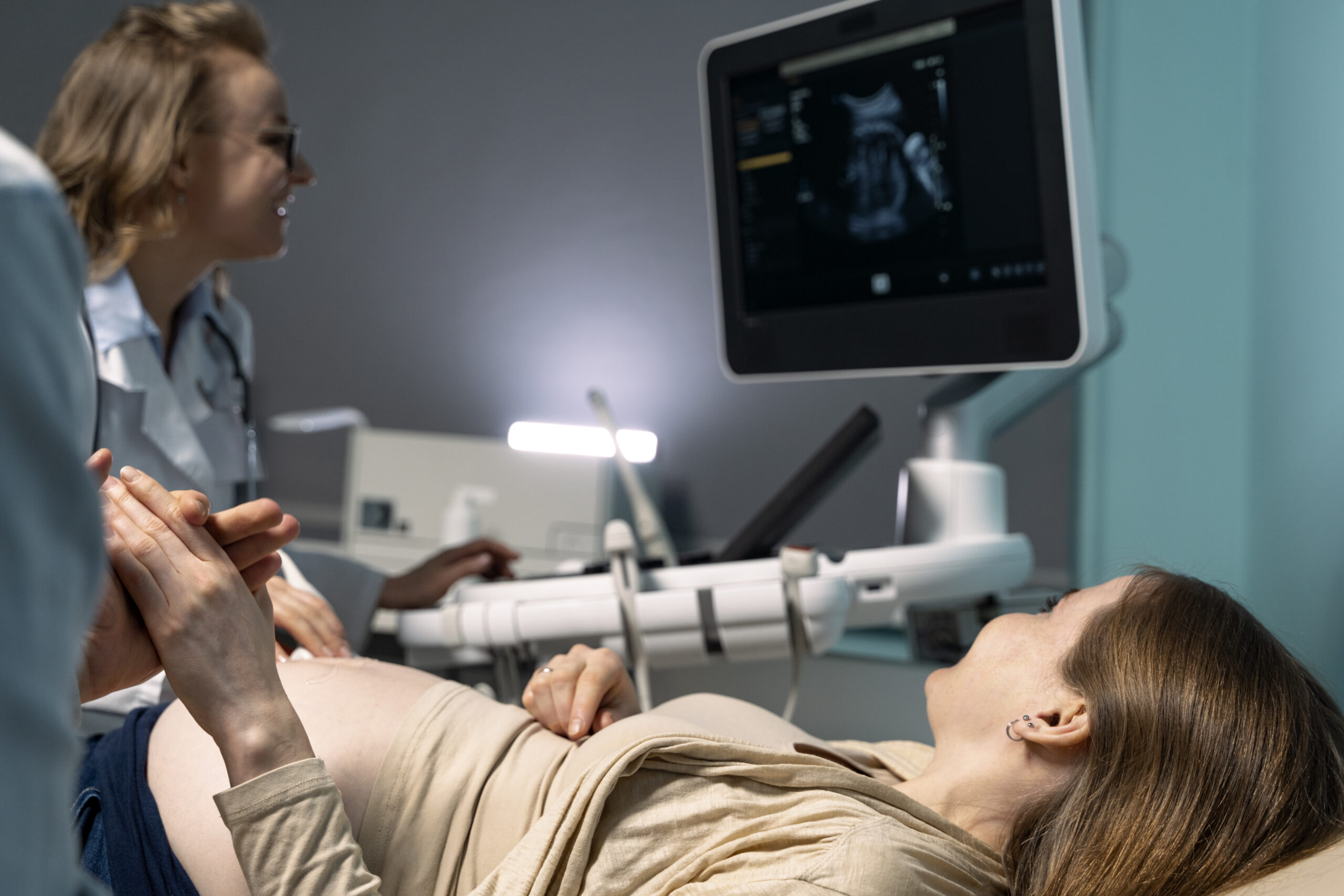
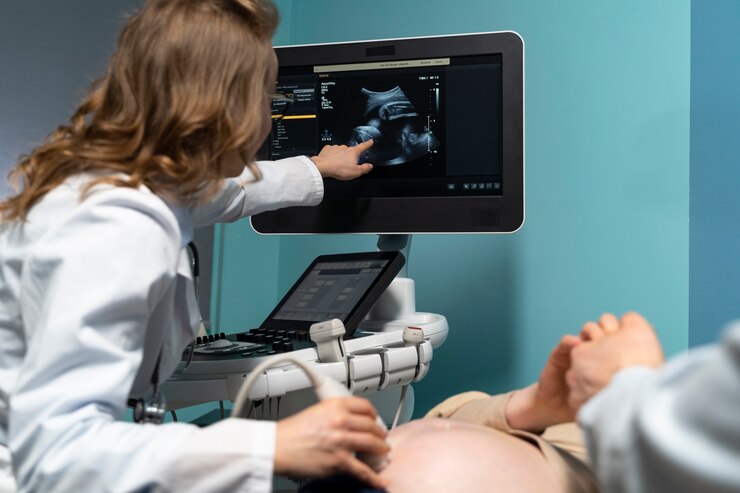
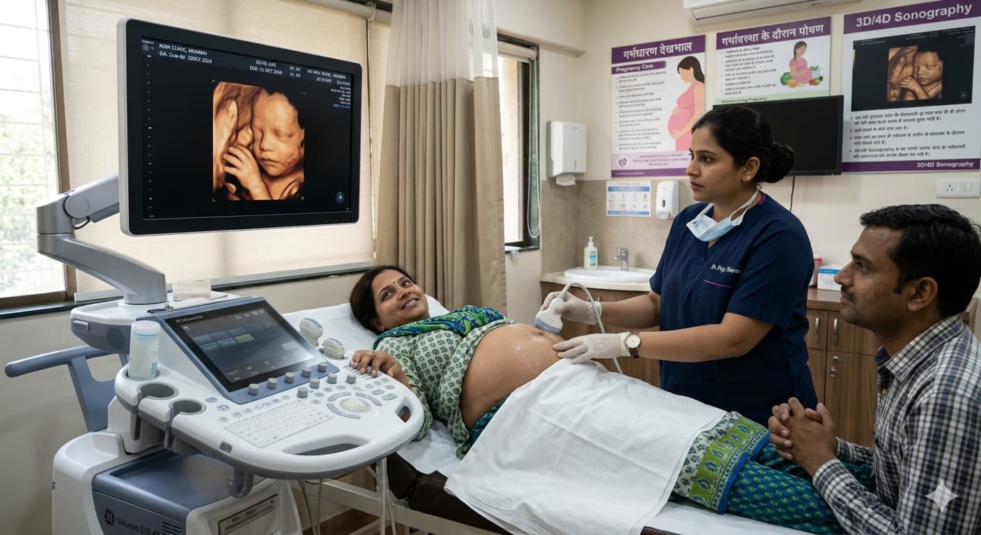
Obstetrics and Gynecology
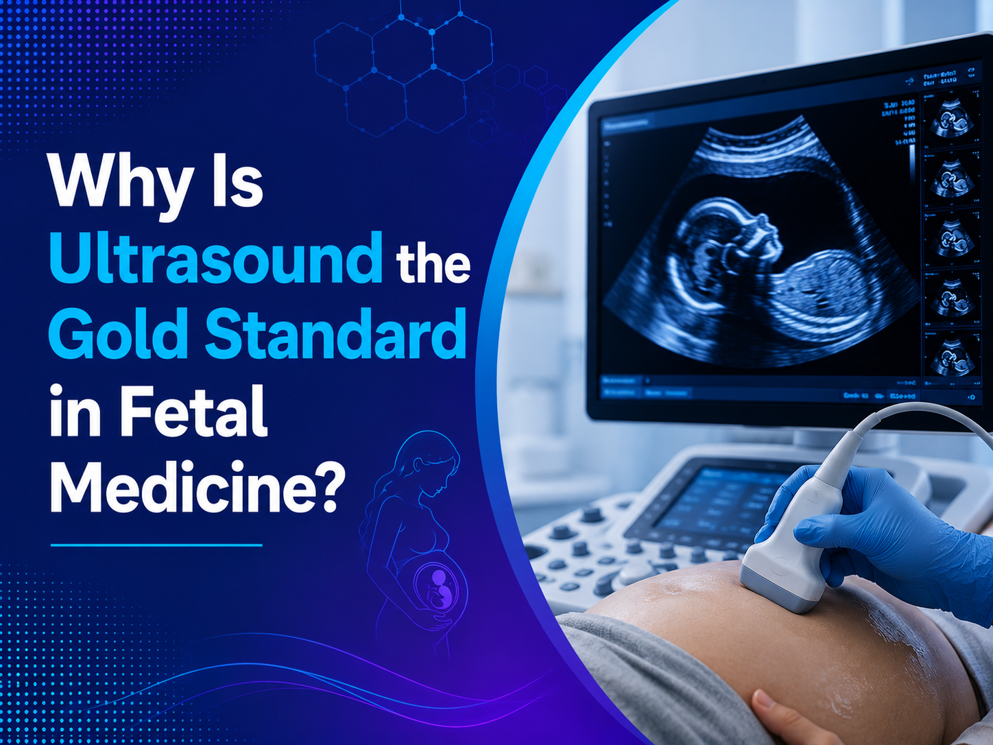
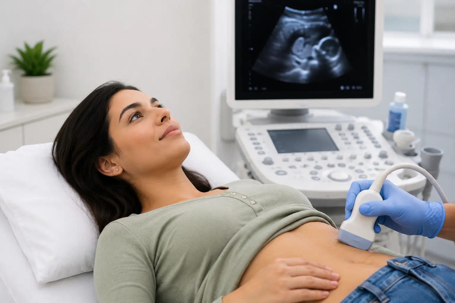
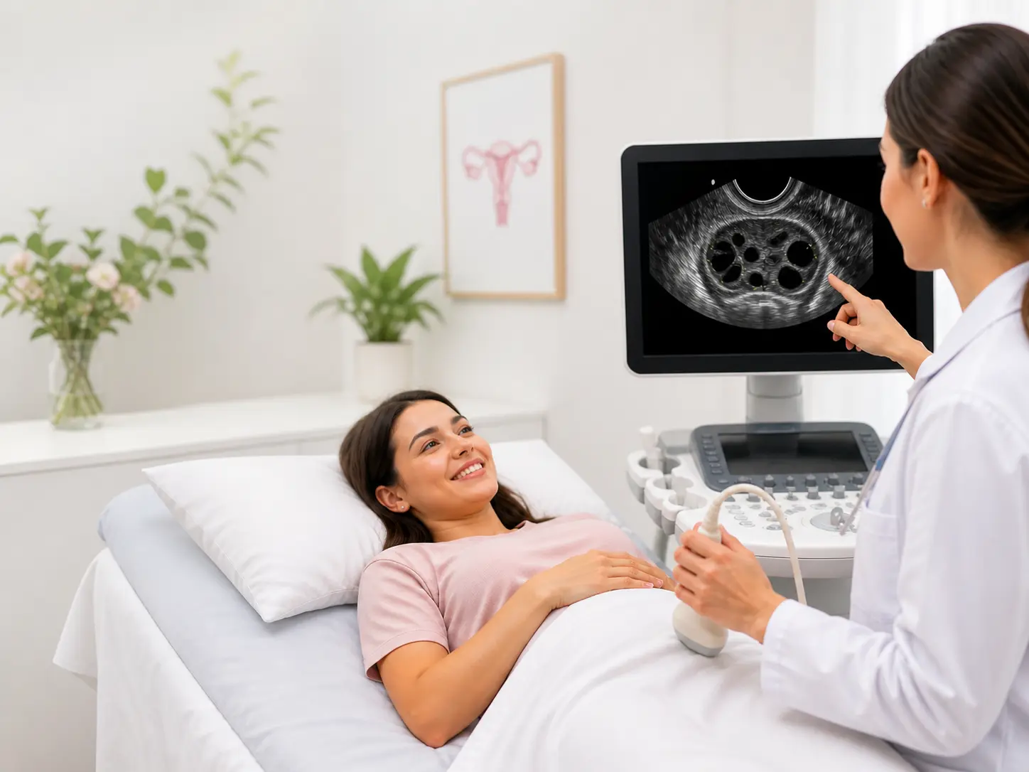
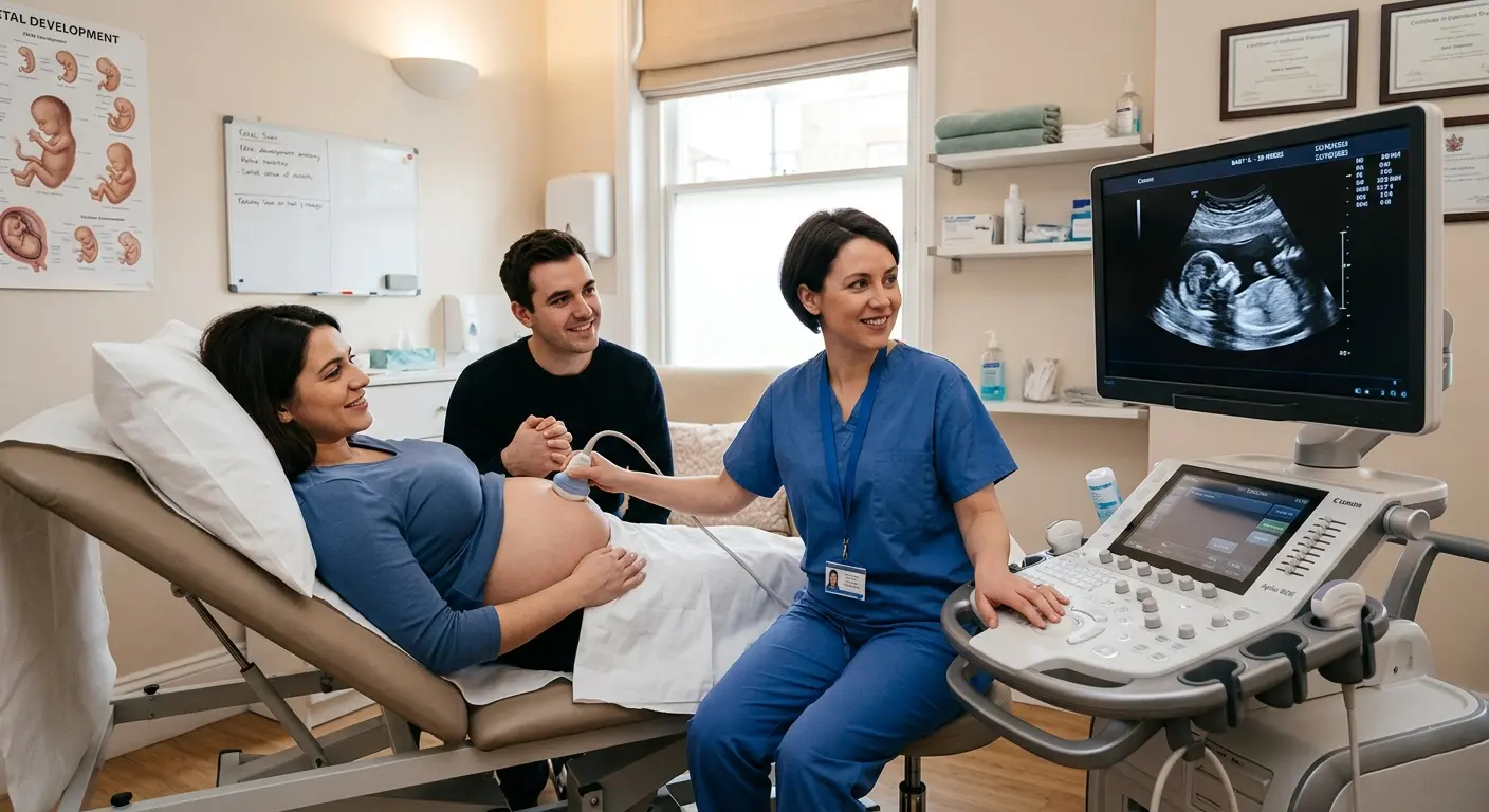
Sonography is important in Prenatal and Gynaecology, which provides vital information on fetal development, pregnancy complications and Gynecological disorders. Obstetric ultrasound provide physicians with a precisely accurate way of monitoring the growth and well-being of the fetus, detecting congenital anomalies, examining the placenta and the amniotic fluid levels and determining the position of the baby in the womb. In gynecology ultrasound imaging plays a significant role in detecting illnesses including fibroid tumors, ovarian cysts, endometriosis, and pelvic inflammatory disease.
Cardiology
On the other hand, echocardiography, a sort of ultrasound imaging tool focusing on the heart, is one of the popular examination methods in cardiology to investigate structure and function of the heart. For instance, this technology allows for close consideration of the walls of the chambers, as well as the valves, clarifying the diagnosis of different cardiac conditions. Echocardiography plays an essential role in the detection of congenital or acquired heart diseases, determination of the functioning of the heart muscles, valve disorders diagnosis, and disease progression monitoring.
Abdominal Imaging
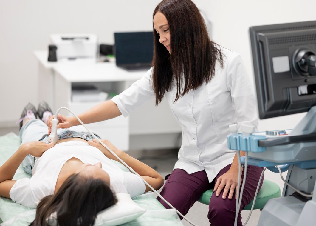
Ultrasonography is the most frequent imaging technique and it is utilized to assess the presence or the absence of any abnormality in the abdominal organs, such as liver, gallbladder, pancreas, spleen, kidneys, and abdominal aorta. It is an effective aid in diagnosing conditions of liver space filling e.g. partly liver cirrhosis, gallstones, pancreatitis cases, renal cysts and abdominal masses. Not forgetting, ultrasound-guided procedures may be done commonly for picking tissue samples or releasing fluid pressure.
Musculoskeletal System
Today, ultrasound finds more and more applications for musculoskeletal imaging to delve into joints and tendons, bean around tendons and scree across the structures of soft tissue. It is useful for the order finding of injuries, tears, inflammation, and other musculoskeletal distortions. Along with these, needle-based therapies including administering joint pain corticosteroids or performing tendon injuries using platelet-rich plasma are also commonly employed.
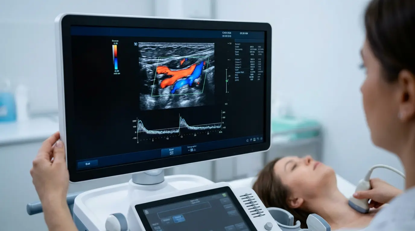
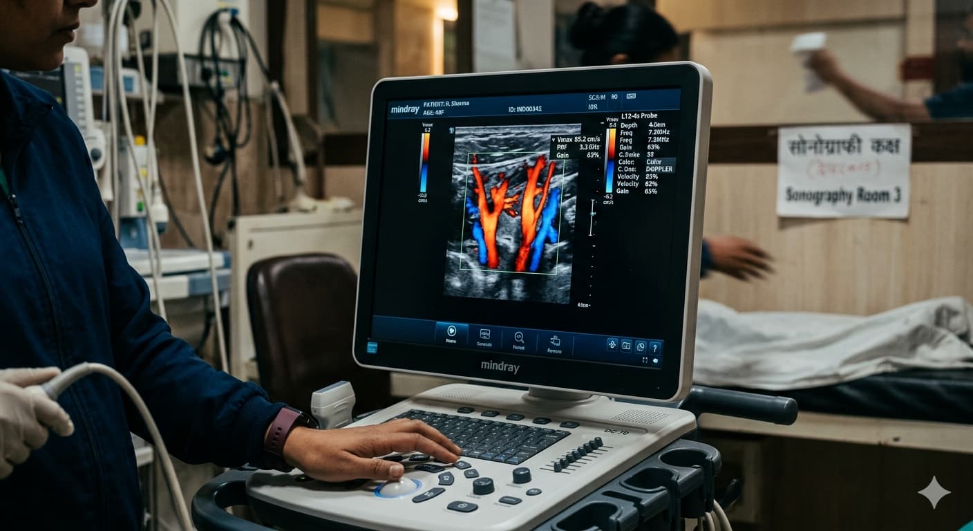
Vascular Sonography
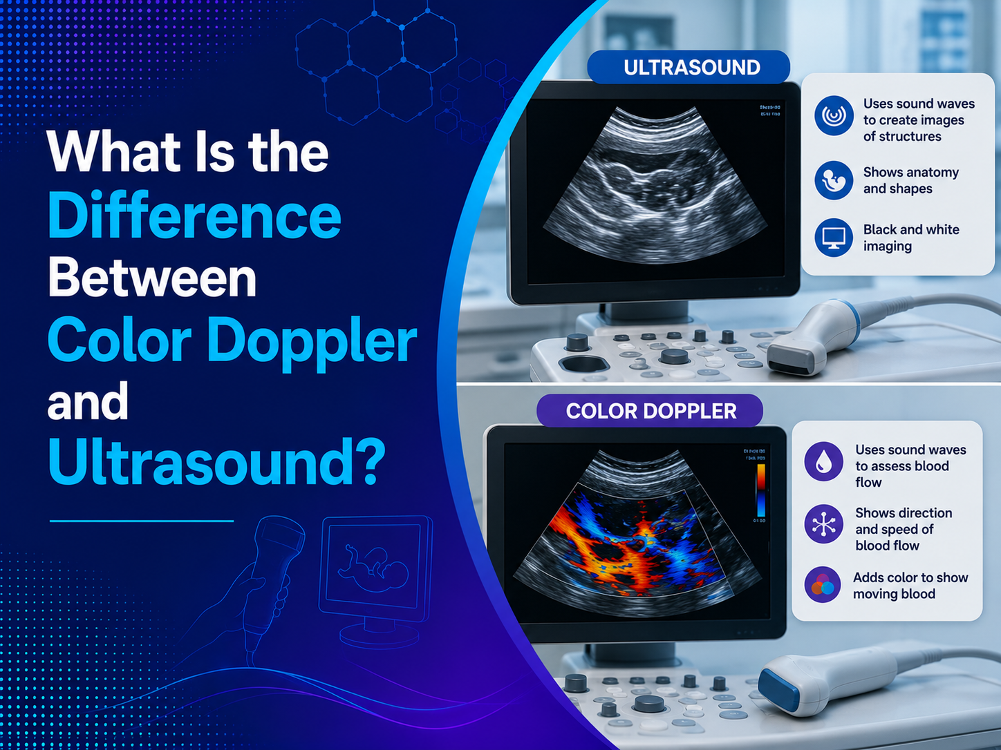
Vascular ultrasound is deployed to visualize the blood flow as well as intactness of blood vessels all over the body. It assists in diagnosing of arterial and venous conditions, e.g. peripheral artery disease, deep vein thrombosis and carotid artery stenosis. Doppler ultrasound is a valuable aid in the assessment of blood flow velocity, obstruction or vessel narrowing and also for guiding endovascular procedures like angioplasty and stent implantation.
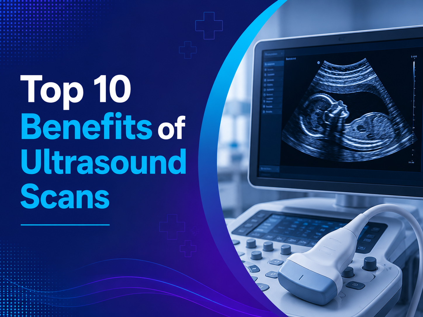
Benefits of Sonography
Sonography has multiple advantages over other modalities of imaging and as such opens up new possibilities for medical diagnosis. One of the principal positive sides of sonography is its excellent safety feature because it excludes any radiation rate and is therefore applicable for all age groups and pregnant women and newborns.
Although sonography may be sometimes painful or uncomfortable, it does not cause these problems as it does not involve any incisions or needles. In vivo visualization abilities allow doctors view the whole types of dynamic information such as fetal motions or blood flow at the same time. In addition to this, the ultrasound devices of current era are compact in size and can be easily transported, which makes point of care imaging possible in various clinical set-ups. Interestingly, ultrasound exams are normally less elaborate and precise when compared to other forms of imaging modalities thus making them more affordable to the patients. Since relying on these benefits help to increase awareness about the use of sonography method for diagnostic purposes among health institutions all over the world.
Sonography offers several advantages over other imaging modalities, making it an indispensable tool in medical diagnostics:
– Safety:
Ultrasound imaging is a modality that has become well established and is generally recognized as a safe option since it does not involve ionizing radiation which is sometimes used with X-rays and CT scans. On the contrary, ultrasound method has employed high frequency sound waves to record the picture of internal tissues. Absence of radiation also helps a eliminate a risk of radiation exposure in both patients and medical professionals, hence, ultrasound equipment is a preferable imaging technique both for pregnant women or children. With the unwanted side effects of aution radiation avoided, ultrasound exams can be repeated as many times as necessary without worries over cumulative exposure, hence, assurance of patient safety and peace of mind.
– Non-invasive:
Ultrasound imaging, as well as its unique characteristic, is its absolute non-invasiveness. Unlike other procedures such as surgery or invasive diagnostic tests, ultrasound exams are often painless and non-incisive, meaning that there is no need for sharp accessories like needles required beforehand. Patients should not bear the pain during the process except the then pressure of the ultrasound probe on the skin. So, there is the absence of anesthesia, or sedation, and the recovery time vanishes. Patients do not have to wait until the exam is over, as it only takes a few minutes, and then they can carry on with their usual lives, without any limits regarding activity or care after the procedure. This extremely comfortable and health associated risks cutting approach makes ultrasound a great choice because it has the lowest risk of complications and infection.
– Real-time Imaging:
Specificity of ultrasound because of its real-time imaging is definitely the strongest feature of this modality. Unlike the pictured images from other modality images, ultrasound offers instant visual feedback that is very beneficial as a healthcare provider will be able to see the motion changes over time. Moreover, the ability to let the visualization happen in real-time is very significant in obstetrics. It helps in showing the fetal movements, cardiac contractions and flow pattern of blood. Furthermore, real-time imaging allows the medical technicians to achieve the confirmation of the position in the procedures that require interventional methods, like ultrasound guided biopsies or injections. This is carried out by depicting dynamic anatomical and physiological discernments as they advance which is crucial for precise diagnosis and treatment planning, resulting in improved outcomes for the patient and possible procedural safety.
– Portability:
Modern day ultrasonography units are notable for their portability and compactness thus being a key feature of bedside imaging. This is in stark contrast to the cumbersome imaging devices that need dedicated facilities and which can be communicated to different related health care settings like hospitals, clinics, physician offices and even remote locations. One portability advantage of ultrasound is that a healthcare provider can conduct the ultrasound exams bedside, in the emergency room, or at a mobile healthcare site. Through the portability of the imaging equipments an ultrasound bridges the gap of accessibility to diagnostic services especially to those living in the margin of the communities or limited resources Portable Ultrasound devices not only provide immediate assessment but also guide the clinicians towards the right decision making, aiding in timely interventions and finally to better patient outcome, especially in Emergency and Critical care situations
– Cost-effectiveness:
Ultrasound examination was recognized as the most cost-effective way of comparing it other imaging techniques like MRI or CT scans. Economical aspect of ultrasound includes cheapness of equipment, consumables, and their maintenance what reduces the prices for healthcare and agricultural facilities and patients. Plus, most of the ultrasound procedures are shorter than other imaging methods, saving both in terms of the time and money for the equipment and human resources. Moreover, the non-invasive characteristics of ultrasound operation makes it possible to do the procedure without medication before, under anesthesia and with monitoring in the post-operational period, and as a result, it reduces related healthcare spending. The money-saving feature makes it a relatively favorable option to be employed both for the mass screening and diagnostic purposes. Such kind of availability of high-quality visualization services to patients regardless of their socioeconomic status improves the society in an economic way. In sum, because the price is balanced to the quality of the service, ultrasound exams contribute to the overall cost containment efforts while preserving diagnostic accuracy and patient satisfaction.
Technological Advancements in Sonography
The latest technological improvements in the range of the capabilities of ultrasound scanning ensure better images, higher processing speeds, and improved accuracy.Some notable innovations include:
– High-Frequency Transducers : The advent of highly sophisticated transducers, which generate high-resolution images, has made it possible to visualize tiny structures and very little lesions distinctly.
–Doppler Ultrasound : Doppler ultrasound tracks the direction and velocity of the blood flow, which is helpful in diagnosing the forms of vascular diseases like atherosclerotic stenosis, venous thrombosis or vascular malformations.
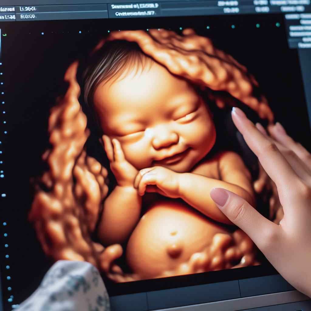
– 3D/4D Imaging: Three- and four-dimensional ultrasound (3D/4D) imaging present real-time reconstructions of incorporated anatomic structures, and afford comprehensive scope of structures and orientation in space.
– Contrast-Enhanced Ultrasound: The contrast agents’ role is to improve the imaging of the vascular structures and the tissue perfusion, specially in the situations when the standard ultrasound imaging is limited.
– Elastography: Elastography is a technique that measures stiffness or the elasticity of tissues, thus providing us with further information about the mechanical properties of organs and aiding the diagnosis of liver fibrosis, breast lesions, and thyroid nodules.
Limitations and Considerations
While sonography offers numerous benefits, it also has some limitations and considerations to be aware of:
– Operator Dependency: Sonography is highly reliant on the proficiency of the practitioners, in the techniques of image acquisition and analysing of the images. Down to operator experience and skillfulness, ultrasound scans can be the key contributor to the quality and diagnostic significance of the study.
– Limited Penetration: Ultrasound rays will mainly penetrate into shallow structures and therefore, imaging of deep-set structures or patients with obesity will be difficult or even impossible.
– Acoustic Shadowing: The existence of shadow is explained by bone, gas or air that acts as an obstacle thus it can block the view of the adjacent organs, hence potentially may decrease the image quality.
– Patient Factors: Image quality and diagnostic accuracy can be affected by the medical conditions of patients, which can include various aspects such as body habitus, bowel gas, and respiratory movement.
Future Perspectives
The future of sonography seems to promise, with the researchers focusing on the development of imaging technologies, proficiency of diagnostic expertise and orientation to clinical applications. Emerging trends in the field of ultrasound include:Emerging trends in the field of ultrasound include:
– Artificial Intelligence (AI): AI algorithms are invented for the automation of the image interpretation, measurements conduct, and improvement of the workflow efficiency.
– Miniaturization: The simpler hand-held ultrasound machines are getting more and more introduced for the point-of-care diagnosis and telemedicine flexibility use.
– Therapeutic Ultrasound: Therapeutic ultrasound methods including high intensity focused ultrasound and US guided ablation are becoming subjects of research today as a non-invasive therapeutic tool for treatment of tumors and pain management, and targeted drug delivery.
Conclusion
Sonography has become an essential diagnostic tool for a wide range of medical disciplines, providing a safe, non-invasive, and adaptable way to view interior organs and structures. Abdominal imaging, musculoskeletal assessment, vascular evaluation, cardiology, and obstetrics and gynecology are just a few of the specialties where ultrasound imaging is essential for diagnosing disorders, directing interventions, and tracking treatment outcomes. Sonography has a bright future ahead of it, with continued research and technology improvements promising to further improve diagnostic
At Ultrascan Diagnostics, we’re dedicated to giving our patients top-notch ultrasound services. We do this by utilizing state-of-the-art equipment and knowledge to guarantee precise diagnosis and individualized treatment. Our team of skilled experts is here to assist with your healthcare requirements, whether you need a basic screening or a thorough diagnostic evaluation.
Take the first step towards better health by scheduling your ultrasound appointment with Ultrascan Diagnostics today.
Faq’s
Are there any risks associated with sonography?
Sonography is considered a safe imaging modality with minimal risks. Unlike X-rays or CT scans, it doesn’t involve ionizing radiation, making it suitable for pregnant women and children. However, as with any medical procedure, there may be rare instances of allergic reactions to ultrasound gel or discomfort during the examination. Your healthcare provider will discuss any potential risks with you beforehand.
How long does a sonography procedure typically take?
The duration of a sonography procedure varies depending on the area being examined and the complexity of the study. In most cases, a standard sonogram lasts between 20 to 60 minutes. However, more detailed examinations or procedures may take longer. Your healthcare provider will give you an estimate of the duration based on your specific needs.
Will I receive the results immediately after the sonography?
In many cases, the sonographer can provide preliminary findings immediately after the procedure. However, a radiologist or physician will review the images more thoroughly to make a final diagnosis. Depending on the facility’s policies and the urgency of the situation, you may receive the results during the same visit or at a follow-up appointment.
How can I schedule a sonography appointment with UltraScan Diagnostics?
Scheduling a sonography appointment with UltraScan Diagnostics is simple. You can call our friendly staff during business hours to book an appointment at a convenient location. Alternatively, you may also be able to schedule online through our website’s appointment booking system. Be sure to have any relevant referral forms or insurance information ready when you call or book online.