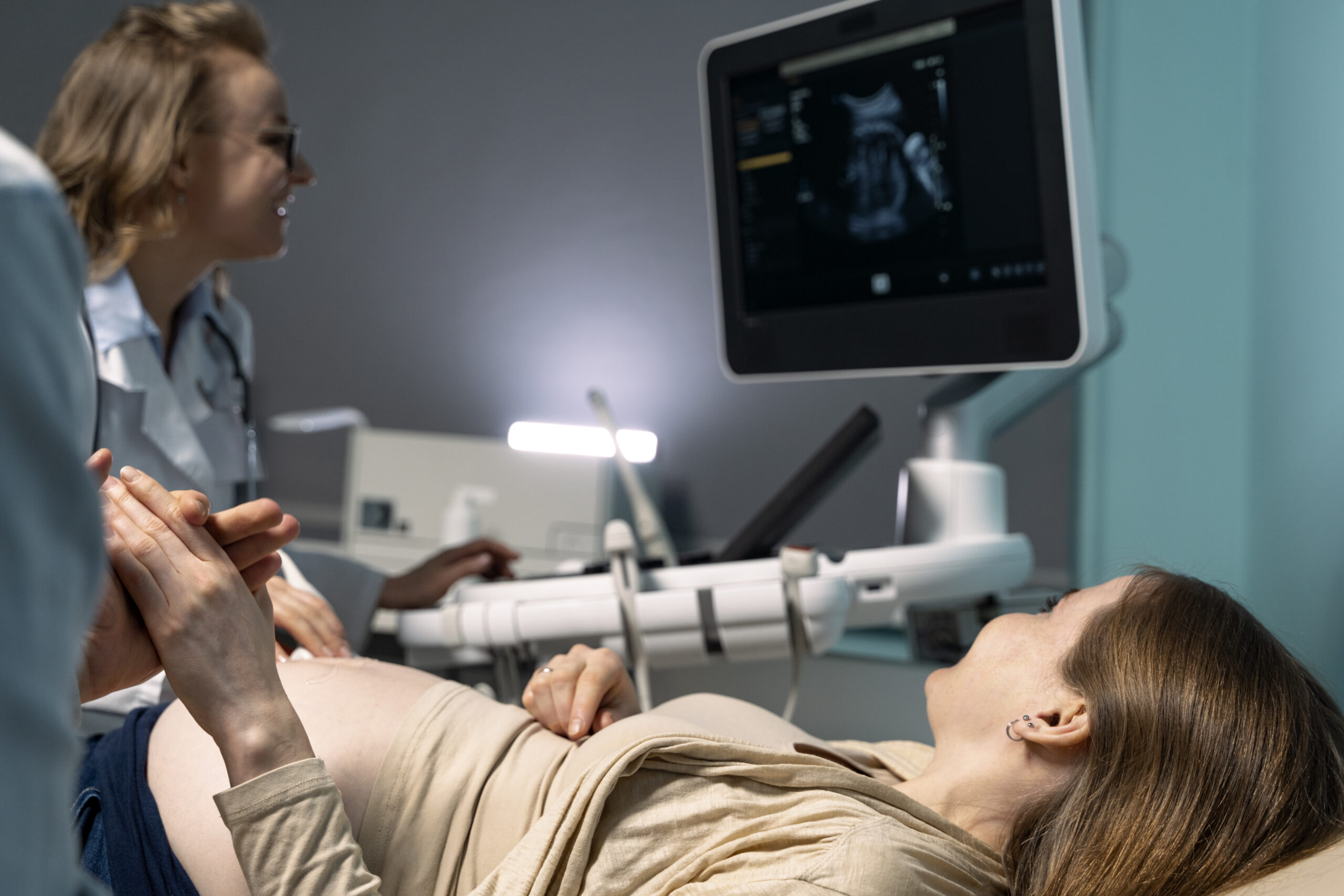
Pregnancy is one of the most exciting – and at times overwhelming – journeys a woman can experience. From the moment you see that positive test, questions begin: What do I eat? Which doctor should I see? And perhaps most immediately, when should you schedule your first ultrasound during pregnancy?
The answer matters more than many first-time mothers realise. Your first ultrasound is not just a moment to see your baby on screen for the first time – it is a medically important scan that provides critical information about your pregnancy, your health, and your baby’s development.
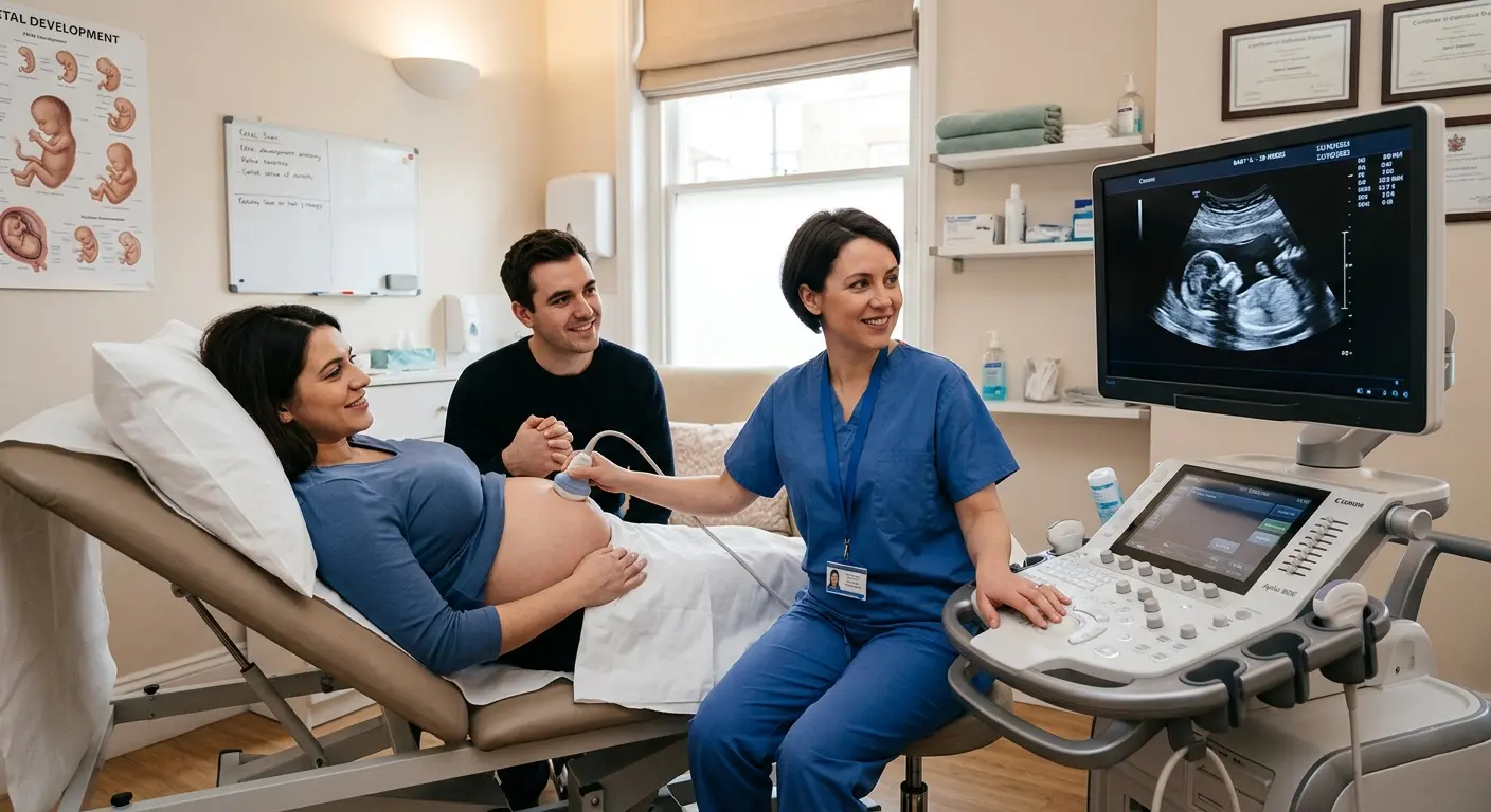
At Ultrascan Diagnostics, your trusted ultrasound diagnostic centre in Indore, we guide expecting mothers through every stage of pregnancy imaging – from the very first scan to the final growth check before delivery. This complete guide tells you everything you need to know, in plain language.
Why Your First Pregnancy Ultrasound Is So Important
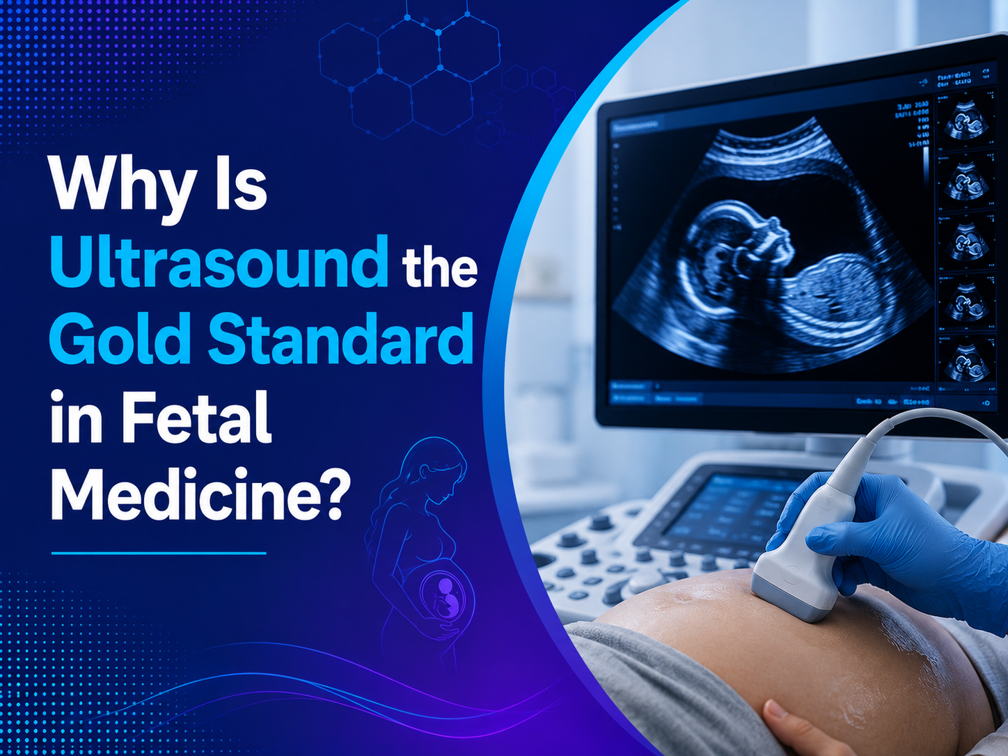
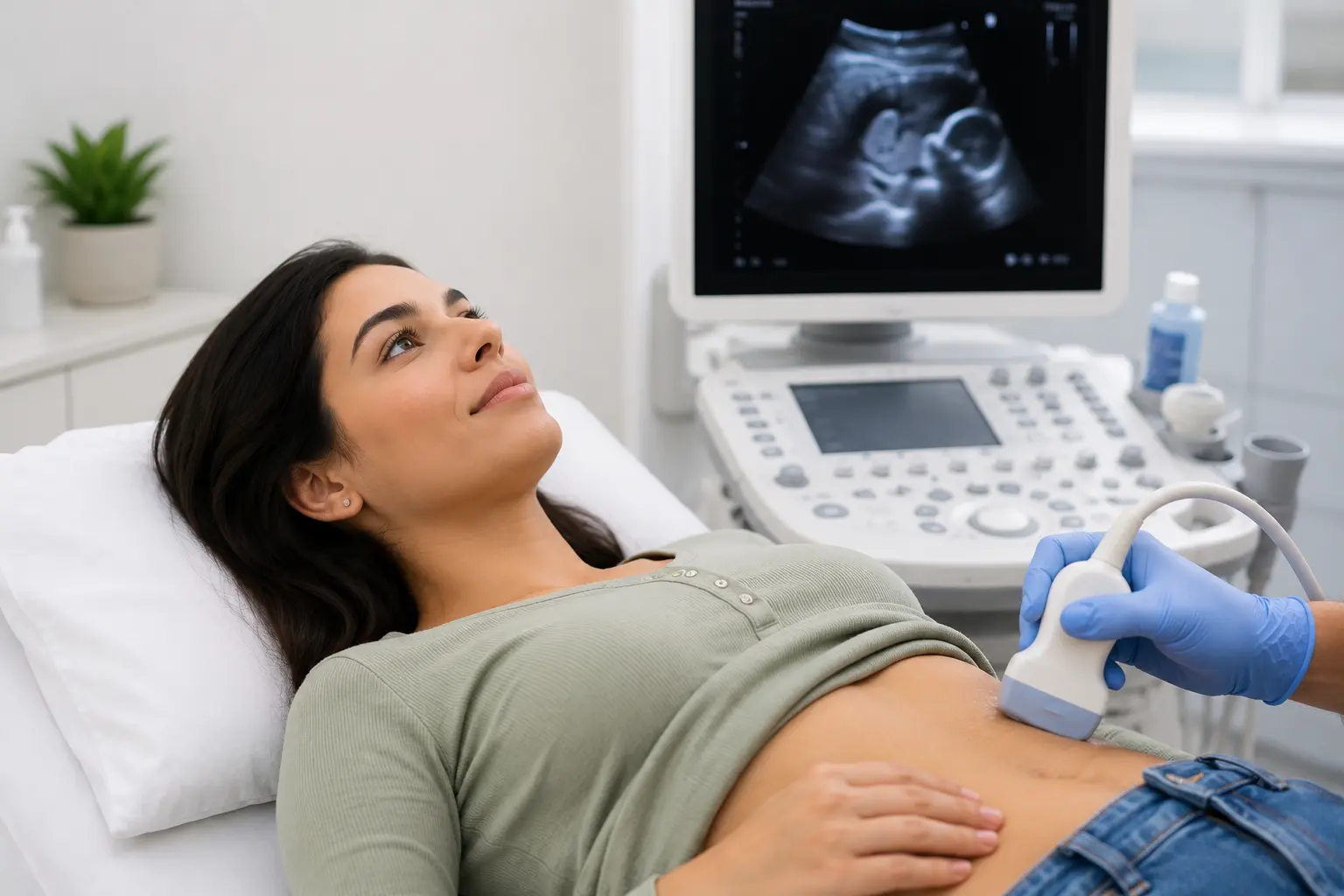
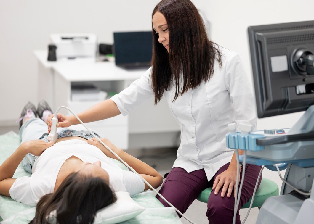
An ultrasound uses sound waves – not radiation – to create real-time images of the inside of your womb. It is completely safe for both you and your baby and can be repeated as many times as clinically needed throughout your pregnancy.
Your first pregnancy ultrasound does several things at once. It confirms that the pregnancy is progressing inside the uterus (not in the fallopian tube, which would be an ectopic pregnancy – a serious condition). It also establishes how many weeks pregnant you are, confirms the number of babies, and checks for early signs of any complications.
Without this scan, your doctor is essentially navigating blind. With it, they have the information needed to plan the right care for you and your baby right from the start.
When Should You Schedule Your First Ultrasound During Pregnancy? The Recommended Timeline
The most commonly recommended time for a first pregnancy ultrasound is between 6 and 10 weeks of pregnancy. However, the ideal timing depends on your specific situation and what the scan is intended to check.
6 to 8 Weeks – Early Viability Scan:
This is the earliest meaningful pregnancy ultrasound and is typically done if you have experienced bleeding, cramping, a history of miscarriage, fertility treatment, or if your doctor simply wants to confirm the pregnancy is healthy. At this stage, the scan confirms a heartbeat, establishes gestational age, and rules out ectopic pregnancy.
8 to 10 Weeks – Dating Scan:
This scan confirms your due date by measuring the baby from crown to rump (head to bottom). It also checks the number of babies and the structure of the uterus and ovaries. If you are unsure of your last period date, this scan is especially important for accurate dating.
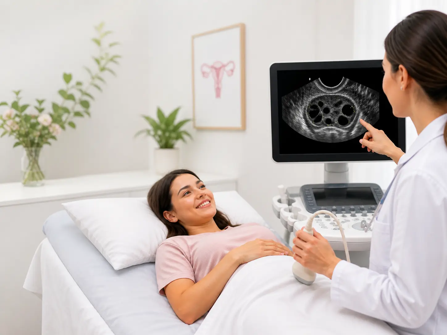
11 to 14 Weeks – NT Scan (Nuchal Translucency Scan):
This is one of the most important scans of the first trimester. The NT scan measures the fluid at the back of the baby’s neck (nuchal translucency), which helps assess the risk of chromosomal conditions such as Down syndrome (Trisomy 21), Edwards syndrome (Trisomy 18), and Patau syndrome (Trisomy 13).
This scan is time-sensitive – it must be done between 11 weeks and 13 weeks 6 days of pregnancy. Missing this window means the opportunity for first-trimester chromosomal screening is lost. According to the NHS, the NT scan combined with a blood test (the combined first trimester screening) gives a much more accurate risk assessment than either test alone.
Pregnancy Ultrasound Schedule After the First Trimester: What Comes Next?
Knowing when to schedule your first ultrasound during pregnancy is just the beginning. A full pregnancy ultrasound schedule typically includes scans at different stages, each with its own purpose.
18 to 20 Weeks – Anomaly Scan (Targeted Imaging / Level II Scan):
This is the most detailed structural scan of the pregnancy. Also called the mid-pregnancy scan, it examines every major organ system of the baby – brain, heart, spine, kidneys, limbs, and face – to check for structural abnormalities. It also confirms placental position and amniotic fluid levels.
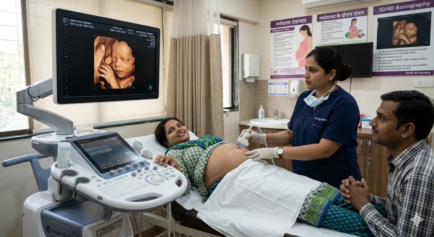
At Ultrascan Diagnostics, we also offer 3D and 4D targeted imaging during this period, giving you a remarkably clear view of your baby’s features and movements in real time. For many parents, this is an unforgettable experience alongside a vital medical assessment.
28 to 32 Weeks – Growth Scan:
A growth scan checks whether the baby is growing at the expected rate for their gestational age. It measures the head, abdomen, and femur (thigh bone) to estimate the baby’s weight. It also evaluates amniotic fluid volume and placental health. Doctors recommend this scan more frequently for high-risk pregnancies.
36 Weeks and Beyond – Late Pregnancy Scan:
In the final weeks, scans assess the baby’s position (head down, breech, or transverse), estimated birth weight, placental grade and position, and umbilical cord blood flow. This information directly informs delivery planning – whether a natural birth or caesarean is safer for mother and baby.
Do High-Risk Pregnancies Need More Frequent Ultrasound Scans?
Yes – significantly more. A high-risk pregnancy requires closer monitoring, and ultrasound diagnostic imaging is the primary tool for this. You may be considered high-risk if you have:
• Twins, triplets, or other multiple pregnancies
• Gestational diabetes or pre-existing diabetes
• High blood pressure or a history of pre-eclampsia
• A history of miscarriage, preterm birth, or stillbirth
• An autoimmune condition, thyroid disorder, or heart disease
• Advanced maternal age (35 years or older)
• IVF or assisted conception pregnancy
• Previous caesarean section
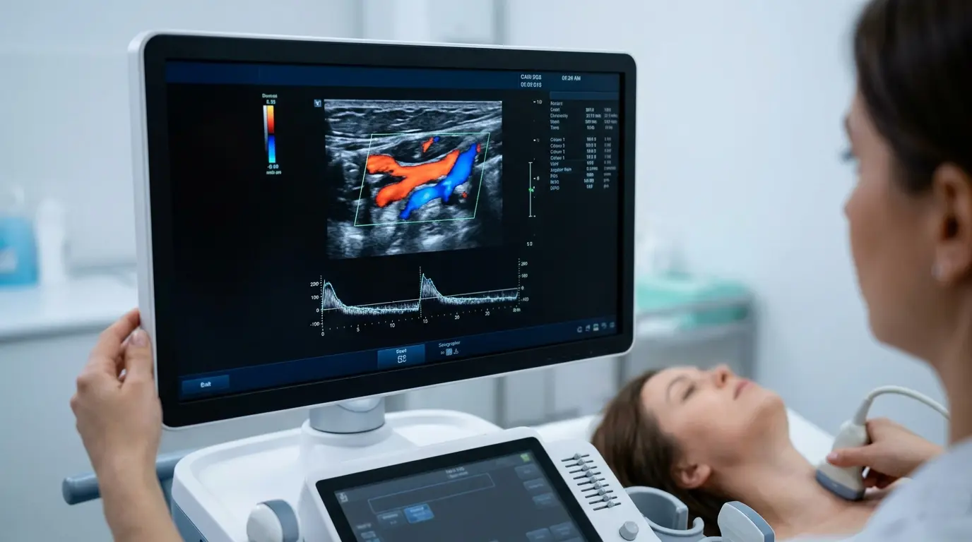
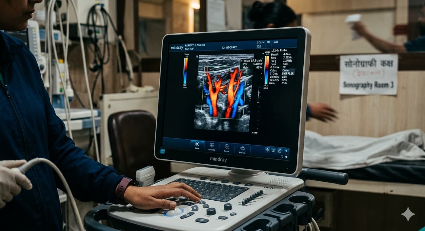
In high-risk cases, your doctor may recommend Colour Doppler scans alongside regular ultrasounds to monitor blood flow through the placenta, umbilical cord, and baby’s brain – ensuring any signs of foetal distress are caught early.
According to the American College of Obstetricians and Gynaecologists, ultrasound is indispensable for managing high-risk pregnancies and should be used as often as clinically indicated to ensure the best outcomes for both mother and baby.
Types of Pregnancy Ultrasound Scans Available at Ultrascan Diagnostics
As a full-service ultrasound diagnostic centre in Indore, we offer every type of pregnancy scan you may need across all trimesters:
• Early Viability Scan (6–8 weeks) – confirms heartbeat and rules out ectopic pregnancy
• Dating Scan (8–10 weeks) – confirms gestational age and due date
• NT Scan / Nuchal Translucency Scan (11–14 weeks) – chromosomal risk assessment
• Early Anomaly Scan (16–18 weeks) – preliminary structural check
• Targeted Anomaly Scan / Level II Scan (18–22 weeks) – detailed structural assessment
• 3D / 4D Ultrasound – detailed imaging of baby’s features and movements
• Fetal Echocardiography – specialised scan of the baby’s heart
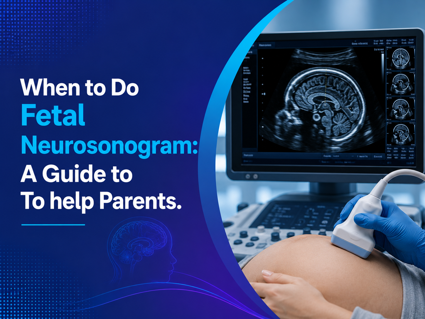
• Fetal Neurosonogram – detailed imaging of the baby’s brain
• Growth Scan (28–32 weeks) – monitors baby’s growth and fluid levels
• Obstetric Colour Doppler – blood flow assessment for high-risk pregnancies
• Late Pregnancy Scan (36+ weeks) – assesses position, weight, and delivery readiness
Every scan at Ultrascan Diagnostics is performed and reviewed by our experienced radiologists, ensuring you receive accurate, timely reports that your obstetrician can act on immediately.
How to Prepare for Your Pregnancy Ultrasound Scan in Indore
Preparation for a pregnancy ultrasound is usually simple, but knowing what to expect helps you arrive relaxed and ready.
• First trimester scans (before 12 weeks): You may need a full bladder, as this helps lift the uterus and provides a clearer image. Drink 2–3 glasses of water about an hour before your scan and avoid emptying your bladder.
• Second and third trimester scans: A full bladder is generally not required, as the baby is large enough to be seen clearly without it. Stay well-hydrated with regular water intake in the days before.
• Fasting: Pregnancy ultrasound scans do not require fasting unless a combined abdominal scan is being performed alongside. Our team will advise you at the time of booking.
• Clothing: Wear loose, comfortable clothing that allows easy access to the abdomen.
• Documents: Bring your prescription, previous scan reports, and any relevant medical records.
When you visit Ultrascan Diagnostics – your nearest ultrasound centre in the Pipliyahana area – our staff will walk you through every step and answer any questions before, during, and after your scan.
Why Expecting Mothers in Indore Choose Ultrascan Diagnostics for Pregnancy Scans
If you are searching for a reliable ultrasound scan in Indore or looking for the nearest ultrasound centre with specialised pregnancy imaging, Ultrascan Diagnostics at Pipliyahana is the name expecting mothers across Indore trust.
• Experienced Specialists: All scans are performed and reported by MD-qualified radiologists with deep expertise in obstetric and gynaecological ultrasound.
• Advanced Technology: Our high-resolution ultrasound machines deliver exceptionally clear images, enabling precise assessment even in early pregnancy.
• Full Obstetric Scan Range: From early viability scans to 3D/4D imaging and fetal echocardiography – everything is available under one roof.
• Comfortable, Private Environment: We understand that pregnancy is an emotional time. Our centre is designed to be calm, clean, and reassuring.
• Timely Reports: We provide prompt reports so your obstetrician or gynaecologist can review your results and plan your care without delay.
• Convenient Location: Situated at Pipliyahana Road, near Kerala Bakery, our centre is easily accessible from across Indore.
• Affordable Packages: Transparent pricing with no hidden charges – because quality prenatal care should be accessible to every family.
The World Health Organization recommends a minimum of eight antenatal contacts during pregnancy, including ultrasound examinations, to reduce complications and ensure safe outcomes for mothers and babies. Ultrascan Diagnostics is your partner for every one of those ultrasound milestones.
Frequently Asked Questions About Pregnancy Ultrasound Scans
Q1. When should you schedule your first ultrasound during pregnancy if you have no symptoms or complications?
Even without any symptoms or concerns, scheduling your first ultrasound between 6 and 10 weeks of pregnancy is strongly recommended. This early scan confirms the pregnancy is in the right location (inside the uterus), verifies a heartbeat, and establishes an accurate gestational age and due date. The earlier you have this information, the better your doctor can plan your prenatal care. If you fall within the 11–14 week window, the NT scan becomes the priority – this is time-sensitive and should not be delayed.
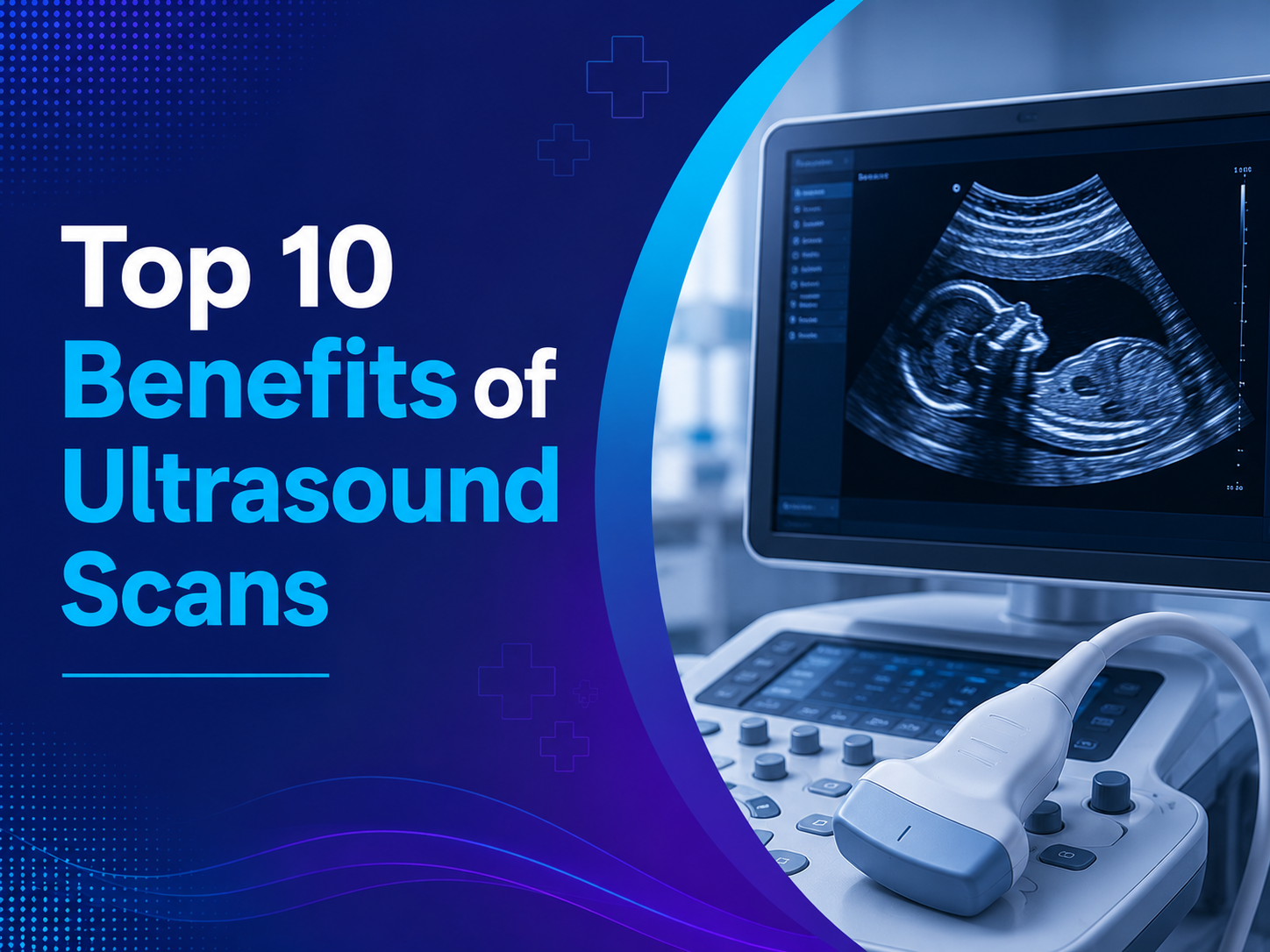
Q2. Is it safe to have multiple ultrasound scans during pregnancy?
Yes – ultrasound scans are among the safest imaging procedures available and pose no known risk to either the mother or the baby. Unlike X-rays or CT scans, ultrasound uses sound waves rather than radiation. Diagnostic ultrasound has been used in obstetrics for several decades and has an excellent safety record across millions of pregnancies worldwide. Your doctor will recommend the number of scans appropriate for your pregnancy, and you can feel confident that each scan is as safe as it is valuable.
Q3. What happens if I miss the NT scan window (11–14 weeks)?
If you miss the NT scan window, first-trimester chromosomal screening through nuchal translucency measurement is no longer possible. However, your doctor has other options. A second-trimester blood test called the Quadruple Marker Screen (or Quad Screen), typically done between 15 and 20 weeks, can still provide information about chromosomal risk. A detailed anomaly scan at 18–20 weeks will also assess the baby’s structural development. While no single test replaces the NT scan at the ideal time, your doctor can still build a meaningful picture of your baby’s health through second-trimester testing. If you are unsure whether you have missed the window, book an appointment immediately – every day counts between weeks 11 and 14.